Open Access
Research Article
Max Screen >>
ISSN: 2348-9790
Copyright: © 2017 Monreal-Pawlowsky T. This is an open-access article distributed under the terms of the Creative Commons Attribution License, which permits unrestricted use, distribution, and reproduction in any medium, provided the original author and source are credited.
Related article at Pubmed, Google Scholar
Over the course of several years, three members of a Bottlenose dolphin (Tursiops truncatus) group showed periodical bouts of pruritus and dermatological lesions. The affected animals were a 25-year-old cow and her 5 and 7-year-old offspring, all housed in a mixed indoor/outdoor facility. The signs were first noted in 2005 and escalated over time until 2012, when a final diagnosis of the problem was attempted.
Diagnostic tests included blood analysis and skin biopsies, swab microbiological cultures from the wheals, intradermal tests and response to treatment with corticosteroids.
Clinical picture and histopathological findings, as well as response to methylprednisolone, were considered compatible with an allergic dermatitis.
Keywords: Bottlenose Dolphins; Dermatological Symptoms; Allergic Dermatitis; Husbandry; Medical Training
Although all mammals should be considered susceptible of developing allergic reactions [1], a search of the existing published literature failed to show any reported cases of allergic disease in cetaceans.
The most common manifestations of allergy in mammals are atopic dermatitis (AD) and respiratory disorders. Allergic respiratory disorders are less frequent in animals than humans, and in fact AD is the most common manifestation of allergy in companion animals. Atopic dermatitis is a chronic inflammatory skin disease that develops as a result of complex interactions between environmental, immunologic and genetic factors [2,3]. In companion animals, the initial diagnosis of AD is made clinically by ruling out other pruritic diseases and following a combination of clinical criteria that are strongly associated with the disease. After clinical diagnosis, laboratory or clinical evaluations such as serum-based or intradermal allergy tests can provide evidence for the allergens involved in AD. However, lack of skin reactivity or allergen-specific IgEs do not rule out the disease [4].
There are no regular features or symptoms that appear in all AD patients, be it humans, or domestic animals such as dogs, cats and horses. However, most common findings include initial onset of signs in young animals up to three years of age, severe pruritus, a species-specific distribution pattern of lesions, corticosteroids responsive pruritus, a chronic-relapsing course and a family history of allergic disease [2,4,5-8].
To the best of our knowledge, there have been no previous reports of allergic disease, or evidence of such, in cetaceans. Similarly, there are no published records of pruritus in Bottlenose dolphins. Here we describe a suspected pruritic allergic reaction to environmental pollen in an adult female Bottlenose dolphin and her two offspring kept under human care.
Over the course of several years, three members of a Bottlenose dolphin group of five animals showed periodical bouts of pruritus and dermatological lesions. These consisted of progressive reddening of the skin (Figure 1) that evolved to macules and then wheals (Figure 2). The affected animals were a 25-year-old cow and her 5 and 7-year-old offspring, housed in a mixed indoor/outdoor facility using filtered natural seawater, located in the middle of a city and surrounded by trees. The outdoor pool was protected against leaves and birds by a net, whereas the roof of the indoor pool had several skylights to provide with natural light, two of which were open and allowed for air as well as pollen to come in at specific dates in springtime.
The clinical signs were more evident in the eldest female. These signs were first noted in 2005 and gradually increased over time until 2012, when a final diagnosis of the problem was attempted.
Onset of symptoms occurred yearly in March or April. In a typical episode, erythema of the prominent ventral areas, such as at the base of the pectoral fins, bilateral to the ventral midline and around the anus and genital area progressed over the course of several days to coalescing macules. In some cases, macules evolved to wheals, some of which presented sloughing of the superficial epithelium (Figure 3). These episodes lasted three to eight weeks, and resolved by May or June. During the episodes, the three animals were seen to rub against the walls of the pools from the onset of symptoms until they remitted.
Diagnostic tests included blood analysis and skin biopsies, swab microbiological cultures from the wheals, intradermal tests and response to treatment with corticosteroids.
Diagnosis of allergic dermatitis followed the classical pruritus diagnostic guidelines, as applied in other mammals such as humans or dogs [2,3,5,8-10]. Cultures proved negative to pathological bacterial and fungal growth. Blood analyses were inconclusive, and only moderate eosinophilia (14% average) was observed. Biopsy samples showed non-specific hyperplasic dermatitis. Intradermal and prick antigen tests were attempted through medical training: to prevent complications such as dry exposure or pressure from an extended stay out of the water, the dolphins were trained to perform the test while in a floating position. Both tests were performed in the ventrolateral abdominal area of the animals. In order to maintain the test area dry, animals were trained to remain in a floating supine position for 30 minutes attended by two trainers, with assistance to breath on command without submerging the test area. One trainer supported the peduncle and the tail, helping it to keep still. A second trainer was placed at the head of the animal, assisting to breath on command. The skin area for testing was prepared by drying and disinfection with clorhexidine at 1% (5% Desinclor® diluted skin preparation, AGB, Madrid, Spain). After the antigen pricks or injections were performed, a silicon cake mould was placed over the test area to prevent small waves wetting the area during the thirty minute waiting time. Neither assay managed to elicit a detectable response to the positive control (histamine).
Standard causes of pruritus such as bacterial or fungal dermatitis, or involvement of parasites were ruled out. Although specific tests for viral pathogens were performed, no histologic lesions suggestive of a viral aetiology were observed. By contrast, response to oral methylprednisolone therapy (Figure 4) showed that it was a corticosteroid-responsive process. The seasonality of the pruritus, wheals, and oedema were other important features suggestive of an allergic process. The seasonal appearance of the clinical signs, coinciding with high pollen content in the area, strongly supported the possibility that these pollens were the most likely cause of the clinical signs.
Palinological records showed that the clinical presentations coincided with the appearance of four pollens, Betula, Pistacia, Celtis and Fagus. Also two out of the seven most common fungal spores, Ganoderma and Drechslera-Helminthosporium, were observed to be present in high quantities during the clinical presentation of disease.
Pruritus, the main clinical sing of allergy, is difficult to observe in aquatic mammals. However, the lesions present in the three animals and the observations made of the three of them rubbing against the walls and bottom of the pool were a key sign to alert of possible pruritus and its relation with the observed lesions.
Clinical picture and histopathological findings were considered compatible with an allergic dermatitis and oral methylprednisolone (Urbason®, Sanofi, Barcelona, Spain) was applied at increasing doses until effect was attained. These treatments proved effective at varying dosages in the three animals, being the highest dose 0,2 mg/Kg once a day (SID) for 10 days, and the lowest 0,02 mg/Kg SID for 3 days.
As is common in atopic dermatitis in other species, the skin was the only affected organ. There is little information, if any, about allergic processes in cetaceans, mainly due to the difficulty of applying the usual diagnostic tools for terrestrial mammals to fully aquatic mammals. Prick tests for instance require of several readings without the puncture sites getting wet. In this case it was aimed to fulfil all the required tests to diagnose an allergic reaction via medical training to avoid placing the animals in unnatural situations, such as out of the water for a prolonged period of time. Training the animals to do all tests voluntarily allowed avoiding management and husbandry problems. Despite all efforts, it was difficult to achieve proper diagnostics such as in domestic non-aquatic animals. Another problem that was encountered is the allegedly low number of affected individuals, since pruritus is very difficult to assess in cetaceans.
The evidence presented in this report was compiled following the classical guidelines for diagnosis in allergy in other mammals. Other causes of dermatosis were ruled out and corticosteroid therapy proved effective at controlling the symptoms. Together with the appearance of the most common symptoms of AD in other mammals, such as pruritus, a specific distribution pattern of lesions, affecting the same areas in the three affected animals, a chronic-relapsing course and a family history of allergic disease, these data are suggestive of an allergic reaction against pollens and spores in a Bottlenose dolphin cow and her two offspring.
Considering the most abundant vegetation in the surrounding area a first attempt of oral vaccination using Parietaria, Platanus, Cupressus and Olea antigens was performed but it has proven unsuccessful so far. Response to an allergy vaccine combining the 4 pollens did not succeed in preventing development of dermatological symptoms over two seasons. At present and after carrying out a detailed study of the most frequent pollination during the critical period, the vaccine composition has been modified (adding Betula and Dactylis and removing Cupressus), but more research is warranted to achieve a positive result.
An in depth revision of literature shows that to date, no allergic disease or allergic reaction have been described in cetaceans. Pruritus or signs of itching in dolphins have not been reported in the scientific literature either.
The authors are aware of an unpublished case of suspected allergic reaction to Plantaina in another dolphin facility that shared similitudes (seasonal presentation, pruritic reaction on ventral areas) with our case (Vicente Arribes, personal communication). It is therefore possible that allergic reactions to environmental antigens are not infrequent and perhaps underdiagnosed in dolphin facilities if the clinical presentation is mild.
The authors would like to thank all the trainers of the Marine Mammal Department because without them, this study would not have been possible.

![]()

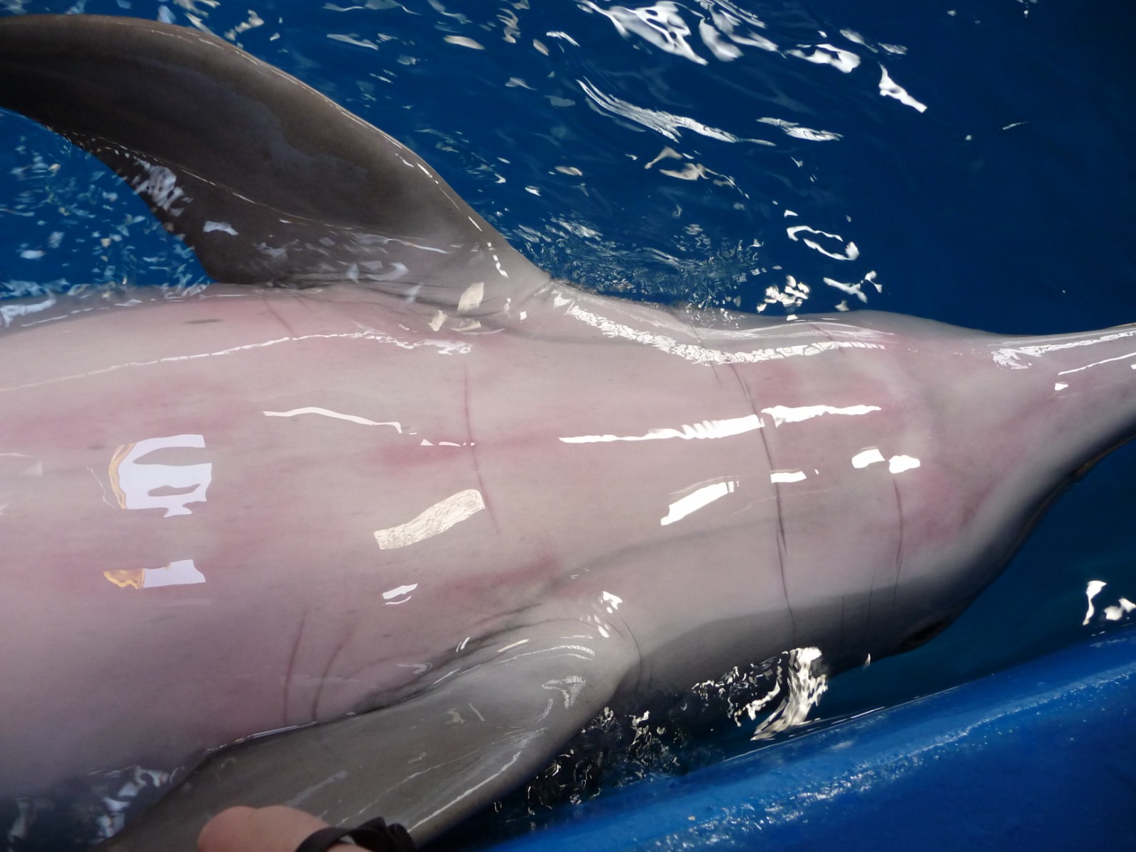 |
Figure 1: Onset of the dermatological problems with reddening of the skin |
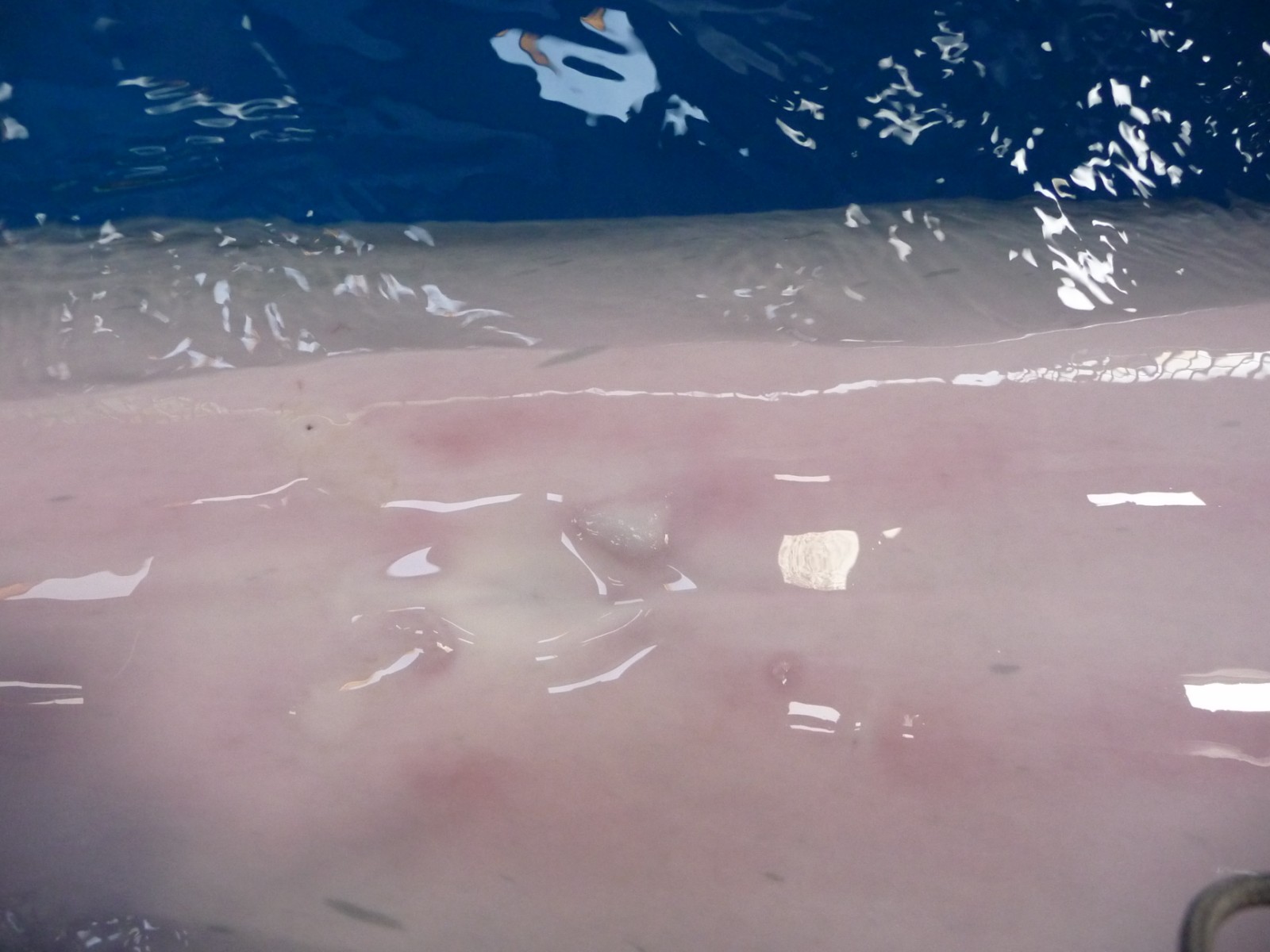 |
Figure 2: Macules and wheals on the ventral area of a bottlenose dolphin |
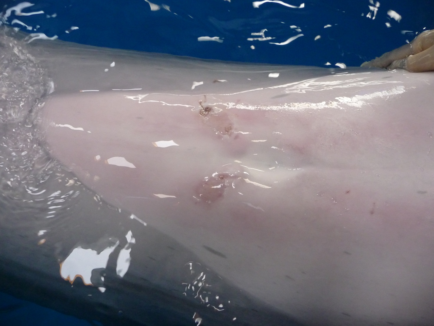 |
Figure 3: Sloughing of skin on the ventral area of a bottlenose dolphin |
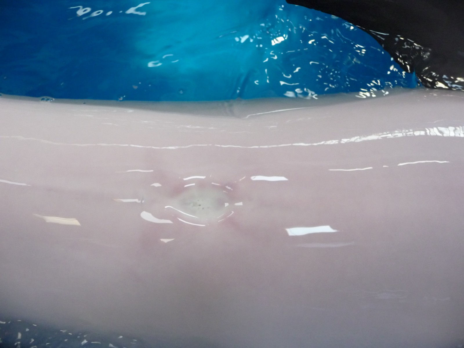 |
Figure 4: Response to methylprednisolone |


