Open Access
Research Article
Max Screen >>
ISSN: 2348-9804
Copyright: © 2021 Ariff S. This is an open-access article distributed under the terms of the Creative Commons Attribution License, which permits unrestricted use, distribution, and reproduction in any medium, provided the original author and source are credited.
Related article at Pubmed, Google Scholar
A simple High-Performance Thin layer chromatography (HPTLC) method has been developed for determination of atenolol in human blood and viscera. The mobile phase was ethyl acetate, acetone, ethyl alcohol, ammonia solution in the ratio of 45:45:7:3. The densitometry scanning at 254 nm was found to be maximum absorption for standard atenolol. The standard and extracted atenolol from samples were detected by HPTLC and confirmed with authenticated spectra at 254 nm. The quantity of the atenolol determined to be 1.6 μg for viscera and 1.5 μg for blood sample.
Keywords:Atenolol; Blood; HPTLC; VisceraChemicals
Atenolol, 4-(2-hydroxy-3-isopropyl-aminopropoxy) phenylacetamide, is a cardioselective β-blocker used for the treatment of cardiovascular disorders, such as hypertension, angina pectoris, cardiac arrhythmias and myocardial infarction [1]. Hypertension is considered as the crucial health issues in majority of the human population leading to heart attack [2]. Increasing blood pressure causes deaths of about nine million people every year, being responsible for 45% of the deaths due to heart diseases and 51% of strokes [3]. Due to the increased population suffering from hypertension every year, the intake of the β-blockers is increased. Since from three decades the β-blockers are used for treating hypertension among which, Atenolol is considered as most prescribed drug for hypertension [2]. In a single treatment of atenolol drug, the systolic and diastolic blood pressure reduces by 15-20% and also lowers the cardiovascular mortality [4]. As a consequence of the increased utilization of these β-blockers and also different case reports suspected as the deaths caused by the β-blockers were published [5-7]. Hence, several methods have been reported for determining atenolol in human plasma and other biological fluids, including high-performance liquid chromatography (HPLC) [8-14], liquid chromatography - tandem mass spectrometry (LC–MS-MS) [15] capillary electrophoresis (CE) [16] and gas chromatography - mass spectrometry (GC–MS) [17-18]. The benefits of these methods include highly sensitive and selective, and some drawbacks could be: expensive equipment, toxic and expensive solvents (mainly HPLC methods) and usually tedious sample pre-treatment when used for analysing biological samples. Therefore, we report a sensitive and specific HPTLC procedure for determining atenolol from the forensic human blood and viscera samples. The advantages of the present method include simple and single step extraction procedure utilizing inexpensive chemicals and short run time. A simple qualitative and quantitative method was required to analyse and detection of atenolol following oral administration. Current procedure showed high variability due to matrix interference. Hardly were any reports on the determination of atenolol by HPTLC method from the forensic samples. In cases like limited blood draw is required or it is provided with the combination of other drugs which cannot be analysed in the whole blood sample, the atenolol analysis in the whole blood could be necessary. Hence, in this paper we report a simple HPTLC method is reported for the determination of atenolol from the forensic samples of human blood and viscera.
Atenolol was of commercial grade procured from local Medical stores, manufactured by IPCA laboratories Ltd (50 mg tablet). All the other reagents were of analytical grade and were obtained from Sigma Aldrich. HPTLC aluminium plates, percolates silica gel-60, t252 (20X20 cm), 0.2 mm thickness purchased from Merck, Germany.
The blood sample and visceral samples were obtained from corpses auto-spaced in the legal Medical Institute (Mandya district hospital) preserved in saturated solution of NaCl at 4 oC until further use.
Stock standard solution of atenolol was prepared in ethanol at 1mg/mL and maintained at -2 oC until and working solution were prepared by diluting with ethanol.
Tissue sample: 2 g of tissue was sliced into small fragments and transferred to 250 ml Erlenmeyer flask, to which 100 mL of chloroform was added, shaken for 5-6 h. The organic layer was separated; the aqueous layer was again extracted with 50 mL chloroform. The chloroform layer was separated; all the two organic layers were pooled and passed through anhydrous sodium sulphate over a funnel, evaporated to dryness and used for HPTLC (for instrumentation purpose organic layer should be passed through Florosil column).
Blood sample: 50 mL of post-mortem blood sample was taken in a 250 ml Erlenmeyer flask to which 50 mL of acetonitrile was added, vortexed for 10 min and incubated for 1 h at room temperature. The two solvent layers were mixed and passed through anhydrous sodium sulphate.
The samples were applied manually in the form of spots on aluminium plates percolated with silica gel-60 F254 (20X10 cm). Space between spots was 20 mm, slit dimension was 0.45 μm and scanning speed was 20 mm/sec. The mobile phase was ethyl acetate, acetone, ethyl alcohol, ammonia solution in the ratio of 45:45:7:3. The linear ascending chromatogram was developed in a glass chamber saturated with mobile phase, till the mobile phase migrate 8 cm. Following the development, the plates were air dried, spots were visualized under UV lamp-254 nm and densitometric scanning was carried by the scanner (200-400 nm range) 3S-M130319 with Wincat software (CAMAG, Switzerland).
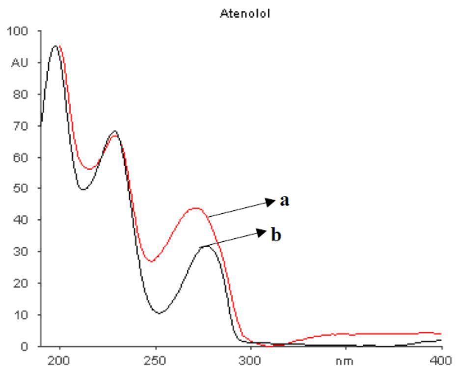
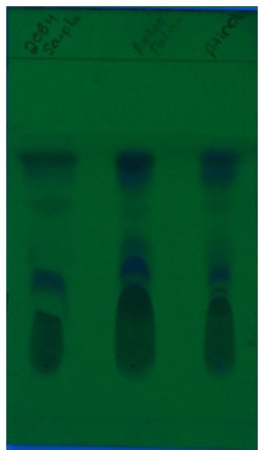
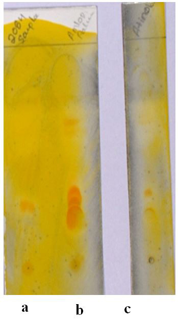
The rate of absorption was found to be 50-60% when its orally administered, and the rest is excreted unchanged in the faces. Atenolol undergoes little or no metabolism and is eliminated primarily by renal excretion [8]. Hence atenolol was found to be stable in blood and viscera samples. Spectra for different concentrations of atenolol (2 to 14 μg/ml) were obtained by scanning at 200 to 400 nm wavelength and optimized the symmetrical peaks of the atenolol for accurate and reproducible results. Standard atenolol peak was compared with the atenolol extracted from the post-mortem viscera (Figure 1) and confirmed the peak and wave length at 254 nm. Atenolol extracted from the blood was compared with the standard atenolol spectra (Figure 2). The HPTLC plate (Figure 3) was visualized under the UV light, the sample extracted from blood (Lane b) and viscera (Lane c) were comparable with the standard atenolol (Lane a). Further these spots were confirmed by using spraying reagent Dragandorf (Figure 4). HPTLC coupled with densitometry was well established and could be the best choice for the identification and analysis of drugs in mixtures and in formulations or from biological samples, due to its high-resolution power. It has been successfully applied for the stability studies and the analysis of a vast number of pharmaceuticals [19]. Different wavelengths have been tested (210, 227 and 254 nm), and found to be at 254 nm and gave good scanning results at which peaks were sharp and symmetrical. Minimum noise and considerable sensitivity of the studied compound was obtained. The TLC coupled with densitometric methods could be applied for the determination of atenolol and in different pharmaceutical formulations with acceptable percentage recoveries [20]. HPTLC technique has been explored for the analysis of various drugs [21-22] and pesticides [23-24]. One of the important applications of HPTLC, includes, several samples could be analysed simultaneously, small quantity of mobile phase, reduces the cost and time of analysis and no restricitons on the choice of solvents and mobile phases [21]. Therefore, these methods do not require any extraction procedure always, and could be developed for drug analysis without any interference from excipients [25].
On comparison with HPLC, TLC does not require pure and high concentrated samples. Highly purified sample are required for HPLC columns as their columns affect separation property. The reported method for atenolol analysis in human whole blood and viscera sample is specific and sensitive. Furthermore, this method may be faster for the analysis as it requires very simple method for the preparation of sample, because of which several number of the analysis can be carried in a single day.
This work provides accurate, sensitive, economic, rapid and selective analytical method for qualitative and quantitative determination of Atenolol from the blood and viscera samples. TLC-Densitometric method for the analysis of atenolol has the advantages over other methods like HPLC, RPHPLC moreover, it can be easily applied in quality control laboratories where economy and time is essential
Director, Directorate of Forensic Science Laboratories, Bangalore and Regional Forensic Science Laboratory, Mysuru are acknowledged for their support.

![]()

 |
|
Figure 1: Densitometry scanning spectra of Atenolol; a: Extracted from Viscera; b: Standard Atenolol |
 |
|
Figure 2: Atenolol extracted from blood sample; a: Compared with the; b: Standard Library |
 |
|
Figure 3: HPTLC plate scanned in UV light at 254 nm; a: Standard Atenolol (2 μg) from Commercially Available Sample; b: Blood Extract of Deceased; c: Visceral Extract of Deceased |
 |
|
Figure 4: Visibility of orange coloured spot when sprayed with Dragandroff reagent; a: Blood Sample; b: Visceral Sample; c: Standard Atenolol |


