Open Access
Research Article
Max Screen >>
ISSN: 2348-9804
Copyright: © 2019 Harel VS. This is an open-access article distributed under the terms of the Creative Commons Attribution License, which permits unrestricted use, distribution, and reproduction in any medium, provided the original author and source are credited.
Related article at Pubmed, Google Scholar
In today’s crime investigation world determination of ABO blood grouping is still a very vital and effective precess in the field of forensic crime scenes. This investigation involves the identification of blood group, on the clothes (Accuse, Victim, injured, Complainer) collected from the scene of crime, and its cross comparision with the blood sample send by medical officer. Hence for this purpose total of 200 cases were included in the study having the samples of blood of accuse, victim, injured, complainer consisting of male and female... The determination of ABO/Rh factor was performed by conventional tube method and matrix gel card. The comparison of both techniques shows a very comparative result. As the red blood cells are sensitized with antibody will get agglutinate in the presence of anti human reagent in the matrix gel card and this will be trapped in the gel column this helps for easy analysis of blood group. However spin tube method is an operator-dependent assay, and is more susceptible to handling errors, the results are not more objective. The matrix gel card method requires Small sample volumes, and gives standardized performance with technical ease, and is with ready automation, and increased biosafety; all these factors have made this technology advantageous. In both techniques the reaction strength for ABO grouping and Rh factor is mainly govern by agglutination reaction intensity between red blood cells and anti-human reagent.
Keywords:Forensic serology; Blood group; Conventional tube technique; Haematology; Matrix gel card
Blood grouping technique is widely used in forensic laboratories for investigation of biological fluids collected from crime scenes. In 1900, Sir Karl Landsteiner discovered the blood grouping technique known as the “ABO” system for which he was awarded with Nobel Prize in 1930 [1]. Edmond Locard a pioneer of forensic science proposed that every criminal carries some trace of evidence with him or her from the scene of crime by which he or she can be linked with the crime. The strongest evidence from the crime scene available is blood or blood stains because the source of blood and their stains help in solving the crime of violence, sexual offences, vehicular accident cases or murder. In cases of natural disasters the prime identification of body part is ABO blood grouping then later comes DNA matching. This is the most commonly followed techniques in today’s forensic laboratory analysis [2,3].
The aim of this study is to determine ABO blood group from the blood samples collected as exhibits from the crime scene and the methods used are spin tube method and matrix gel System. The comparative study for these two method was carried out by testing total 200 samples of blood in the form of clothes or different types of exhibits which having blood strains.
This study is carried out for the comparative study between spin tube method and matrix gel card method for blood group identification on the basis of efficacy sensitivity and specificity [12-14].
1. The Matrix AHG (Coombs) test card:- Matrix forward and reverse grouping card with auto control has six micro-tubes prefilled with auto control a gel in a suitable buffer. The first three micro-tubes contains Monoclonal Anti-A, Anti-B and Anti-D. The forth, five and six number Micro-tubes has neutral gel. The Forth tube is used as negative control. The fifth and sixth are used for reverse grouping.
2. Red Blood cells:- The Fresh blood samples of “A, B and O” blood group were kindly procided by blood bank of J.J. group of Hospitals, Mumbai, Maharashtra, India.Matrix card reader ( Matrix Auto Max-80)
3. Matrix Diluent- LISS:- for three preparation of fresh cells.
4. Gel card centrifuge (Imperial Biotech LLP, Tulip gel card centrifuge)
5. Micropipette
In the conventional spin tube method the ABO forward grouping is determined in the presence of the blood group antigens A, B and O by testing the RBC’s with known antisera, specifically Anti-A, and Anti-B. On the other hand, the ABO reverse grouping method results into the presence of the expected ABO blood group antibodies by testing the serum or plasma with known A1 and B red blood cells. In case of matrix gel test the ABO forward grouping were performed in the Anti-A, Anti-B, micro-tubes, which contain the specific antibody incorporated into the gel.
ABO blood group can be determined from the fresh blood samples by detection of antigens on RBC’S (forward method) or detection of antibodies in serum (reverse method). The blood samples are received in sterile bulb by forensic laboratories from the medical officers. The testing sample is taken into the tube and saline 0.2%, was added to the sample. This mixture was then centrifuged at about 3000 rpm for five minutes. The supernatant was then discarded and the suspended cells were rewashed with saline for two times. Resuspend the button of cells in fresh normal saline. After this the cell suspension was prepared in 2% saline.
In the tile with two cavities added one drop of anti-A serum and anti-B serum in the above marked cavities A and B respectively. Added one drop of about 2% cell suspension in each cavity and mixed the contents thoroughly. The tile was roated for 5 minutes and results were examined macroscopically as well as microscopically for agglutination.
Removed the serum carefully. Marked the cavities of tile as A and B. Added one drop of serum in each cavity. Added one drop of 2% cell suspension of A and B cells in the respective cavities and rotated the tile for 5 minutes results are examined macroscopically as well as microscopically for agglutination.
In this method ABO reverse grouping was performed in the buffer gel micro-tubes. Formation of agglutination indicates the presence of an antigen-antibody reaction, while lack of agglutination indicates the absence of an antigen-antibody reaction. Agglutinated red blood cells are trapped in the gel at various levels within the micro-tube depending on the size of the agglutinates. Free non agglutinated red cells pass through the gel and form a button of red blood cells on the bottom of the micro-tube. The control micro-tube must be negative for the results to be valid. In the gel test, the reagent red blood cells are combined with sample serum/plasma in the upper reaction chamber of the micro-tube of card. Following an incubation period to enhance antigen/antibody interaction, the sensitized red blood cells react with the antibodies incorporated in the gel of the micro-tube during a centrifugation step. Agglutination indicates the presence of an antigen/antibody reaction while lack of agglutination indicates the absence of an antigen/antibody reaction. Agglutinated red blood cells become trapped in the gel at various levels within the micro-tube, depending on the size of the agglutinates. Free non agglutinated red blood cells pass through the gel and form a button of red blood cells on the bottom of the micro-tube.
Prepared 5% of blood cells suspension in matrix diluent LISS, by taking 0.5 mL LISS + 50 μL of whole blood or 25 μL of packed blood cells. Mixed gently and used for forward grouping.
Prepared 0.8% blood of cells suspension in matrix diluent LISS as follows: - Washed fresh A, B cells with 0.9% saline till the supernantant is clear. Add 1 mL of LISS in Each labelled test tubes as A and B. Used for reverse grouping.
Pipetted 10 μL of 5% blood cell suspension in the microtubes 1 to 4 (A-B- -D- Control). Pipetted 50 μL of 0.8% known ‘A’ blood cell suspension in the microtube 5. Pipetted 50 μL of 0.8% Known ‘B’ blood cell suspension in the microtube 6. Pipetted 50 μL of serum of A and B in the microtubes 5 and 6. Allowed the card to incubate for 10 min.at room temperature. The card was removed and the results were recorded.
Agglunation in the forwarding grouping and either haemolysis or Agglunation in the reverse grouping were interpreted as a positive reaction. The results of ABO and Rh type of samples were recorded [15-17].
The reaction strength may be recorded by grading of the Agglutination Reaction Intensities:-
The Red blood cells possesing the corresponding antigen will agglutinate in the presence of specific antibody, and will trapped in the gel column, and gets settle at the bottom of the microtube. The control microtube (Ctrl) must be negative to validate the forwarding results [8-10].
Positive Reaction:- A clear line on the surface of the gel column is formed by the agglutinated blood cells or sometimes it dispersed in the gel column [8-10].
Negative Reaction:- Non Agglutinated red blood cells settle at the bottom of micotube forming a compact button [8-10].
G++++ (G4+):- Agglutinated red blood cells form a line at the top of the gel microtube.
G+++ (G3+):- Most agglutinated red blood cells remain in the upper half of the gel microtube.
G++ (G2+):- Agglutinated red blood cells are observed throughout the length of the microtube. A small button of red blood cells may also be visible at the bottom of the gel microtube.
G+ (G1+):- Most agglutinated red blood cells remain in the lower half of the microtube. A button of cells may also be visible at the bottom of the gel microtube.
G± Most agglutinated red blood cells are in the lower third part of the gel microtube.
Negative: - Negative all the red blood cells pass through and form a compact button at the bottom of the gel microtube.
Agglutinated red blood cells form a line at the top of the gel and non-agglutinated red blood cells form a compact button at the bottom of the gel microtube.
H:- Hemolysis of red blood cells
Out of 200 blood samples (which include Accuse, Victim, Suspect, injured, Deceased, Vitness etc.) cases, 5% (10 out of 200) shows “inconclusive or invalid” results.
Figure 1 shows, G (3+) agglutination reaction in forward A-cells and G (3+) agglutination reaction in reverse grouping B-serum and shows negative reaction for B-cells and A-serum. Hence the blood group is ‘An’ Rh+.
Figure 2 shows, G (4+) agglutination reaction in forward A-cells and G (4+) agglutination reaction in reverse grouping B-serum, and shows negative, reaction for B-cells and A-Serum. Hence the blood group is ‘An’ Rh+”
Figure 3 shows, G (4+) agglutination reaction in forward A-cells and G (2+) agglutination reaction in reverse grouping B-serum, and shows negative, reaction for B-cells and A-Serum. Hence the blood group is ‘An’ Rh+”
Figure 4 shows, G (4+) agglutination reaction in forward B-cells and G (4+) agglutination reaction in reverse grouping A-serum, and shows negative, reaction for A-cells and B-Serum. Hence the blood group is ‘B’ Rh+.
Figure 5 shows, G (4+) agglutination reaction in forward B-cells and G (2+) agglutination reaction in reverse grouping A-serum, and shows negative, reaction for A-cells and B-Serum. Hence the blood group is ‘B’ Rh+.
Figure 6 shows, G(4+) agglutination reaction in forward B-cells and G (1+) agglutination reaction in reverse grouping A-serum, and shows negative, reaction for A-cells and B-Serum. Hence the blood group is ‘B’ Rh+.
Figure 7 shows, G (4+) agglutination reaction in forward A-cells and B-cells and negative agglutination reaction in reverse grouping for A-serum and B-serum. Hence the blood group is ‘AB’ Rh+.
Figure 8 shows, Negative agglutination reaction in forward A-cells and B-cells and G (4+) agglutination reaction in reverse grouping for A-serum and G (3+) agglutination reaction for B-serum. Hence the blood group is ‘O’ Rh+.
Figure 9 shows, Negative agglutination reaction in forward A-cells and B-cells and G (2+) agglutination reaction in reverse grouping for A-serum and B-serum. Hence the blood group is ‘O’ Rh+.
Figure 10 shows, Negative agglutination reaction in forward A-cells and B-cells and G (4+) agglutination reaction in reverse grouping for A-serum and G (2+) agglutination reaction for B-serum. Hence the blood group is ‘O’ Rh+.
Figure 11 shows, Negative agglutination reaction in forward A-cells and B-cells and G (2+) agglutination reaction in reverse grouping for A-serum and B-serum. Hence the blood group is ‘O’ Rh+.
Figure 12 shows, Negative agglutination reaction in forward A-cells and B-cells and G (3+) agglutination reaction in reverse grouping for A-serum and B-serum. Hence the blood group is ‘O’ Rh+.
Figure 13 shows, Negative agglutination reaction in forward A-cells and B-cells and G (2+) agglutination reaction in reverse grouping for A-serum and G (1+) agglutination reaction for B-serum. Hence the blood group is ‘O’ Rh+.
Figure 14 shows, G (4+) agglutination reaction in forward A-cells and B-cells and G (3+) agglutination reaction in reverse grouping for A-serum and B-serum. Hence the blood group is cannot be concluded.
Figure 15 shows, G (2+) agglutination reaction in forward B-cells and negative agglutination reaction A-cells and G (4+) agglutination reaction in reverse grouping for A-serum and B-serum. Hence the blood group is cannot be concluded.
Figure 16 shows, G (4+) agglutination reaction in forward B-cells and negative agglutination reaction A-cells and G (3+) agglutination reaction in reverse grouping for A-serum and negative agglutination reaction for B-serum. Hence the blood group is cannot be concluded
Figure 17 shows, G (4+) agglutination reaction in forward A-cells and B-cells and G (4+) agglutination reaction in reverse grouping for A-serum and B-serum. Hence the blood group is cannot be concluded
This study presents number of advantages of blood grop cross matching by using Matrix gel card method over routine spin tube method. The hemagglutination in positive wells was strong, so that they were easily seen with the naked eye.
The ideal condition for the agglutination reaction for blood group type “A” is Figure 2, for blood group type “B” is Figure 4. For blood group type “AB” is Figure 7, for blood group type “O” is Figure 12. It was observed that Figure 1 shows G (3+), reaction and Figure 3 shows G (2+) reaction, but when these samples were preocessed, the correct reaction for reverse (Serum) grouping was some times observed difficult to conclude. Similarly the results of samples in Figure 5, Figure 7, Figure 14 shows G (2+), G (1+)...etc reaction and but when these samples were preocessed with routine spin tube method, the very poor agglutination reaction was observed.
Figure 15, Figure 16, Figure 17, and Figure 18 shows the inconclusive or invalid results by both routine spin tube method, and Matrix gel card method.
In our study it was observed that 5% (10 samples out of 200) samples which showed invalid/ Haemolysed / Inconclusive results by our routine spin tube method, was giving correct agglutination by gel card technique. Which is coated in the studies by Kaur R, Rumsey DH, and Malyska H. et.al?
It wass observed that Matrix gel card test is better than Spin saline tube method because of its simplicity, stability of results, better handling, long time recorded, dispensation of controls with comparable sensitivity and specificity which is follow with this study (Col et al., 2008).
As the Matrix gel test uses an increased serum to cell ratio and there is no need of wash phase, thus reducing possibility of elution of weakly bound antibodies hence the false positive screens of results were reduced using the matrix gel test system. (Bromilow et al, 1992).
Matrix gel card test results remain stable within the gel, allowing rereading and it can be photocopied. The disposal of plastic cards is also very easy, which increases standardisation of laboratory techniques and introduces more objective reading of agglutination reaction. (Bromilow et al, 1992).
The blood group antigens for ABO and Rh factor were detected upto 12 Months. The number of non-specific anti bodies and false positive sereens of results were reduced using matrix gel system.
From above observations it was concluded that the Matrix gel card technique is more suitable and less time consuming. The results are more stable can be recorded after long time.The test can be carried out with very small sample. In conclusion Matrix gel card test has been shown to be very efficient in forensic fields to get the blood group from the blood samples of Accuse, Victim, injured, Complainer Consisting of Male and Female.
Author thanks to Director General (Legal & Technical) Home Dept. Govt. of Maharashtra and Forensic Science Lab Laboratory, Mumbai, for the facilities to do this analysis.

![]()

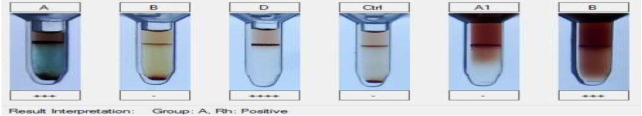 |
Figure 1: G (4+): Rh; G (3+): A-cells, B-Serum; Negative (-): B-cells, Ctrl, A-serum |
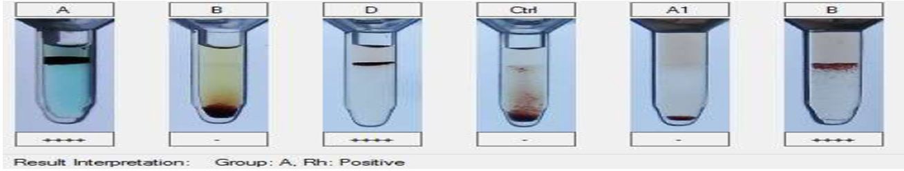 |
Figure 2: G (4+): A-cells, Rh, B-Serum; Negative (-): B-cells, Ctrl, A-serum |
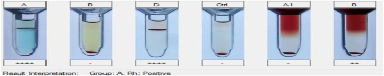 |
Figure 3: G (4+): A-cells, Rh; G (2+): B-Serum; Negative (-): B-cells, Ctrl, A-serum |
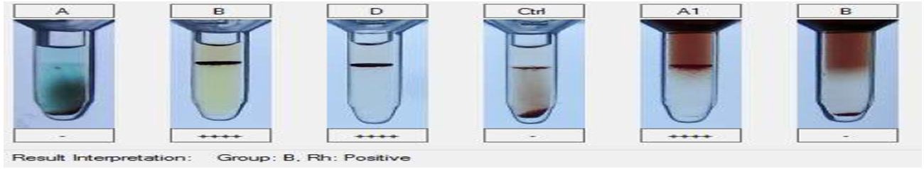 |
Figure 4: G (4+): B-cells, Rh, A-Serum; Negative (-): A-cells, Ctrl, B-serum |
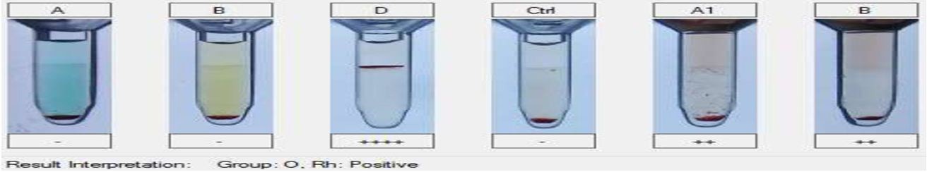 |
Figure 5: G (4+): B cells, Rh; G (2+): A-Serum; Negative (-): A-cells, Ctrl, B-serum |
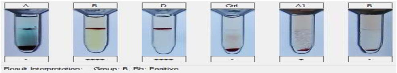 |
Figure 6:G (4+): B-cells, Rh; G (1+): A-Serum; Negative (-): A-cells, Ctrl, B-serum |
 |
Figure 7:G (4+): A-cells, B-cells, Rh; Negative (-): A-serum, B-Serum, Ctrl |
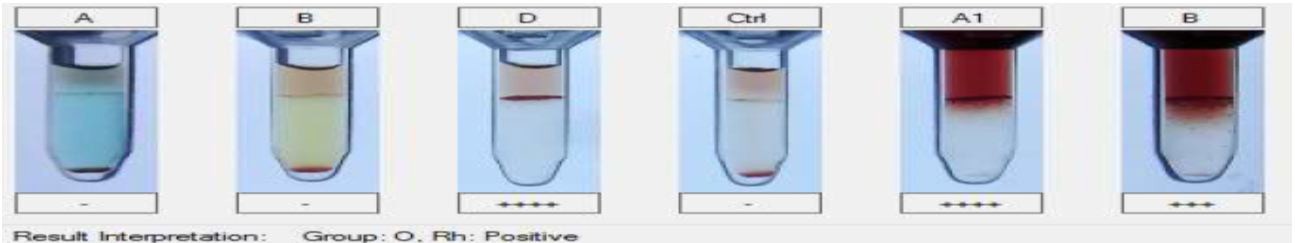 |
Figure 8: G (4+): Rh, A-serum; G (3+): B-Serum; Negative (-): A-cells, B-cells, Ctrl |
 |
Figure 2: G (4+): Rh; G (2+): A-serum, B-Serum; Negative (-): A-cells, B-cells, Ctrl |
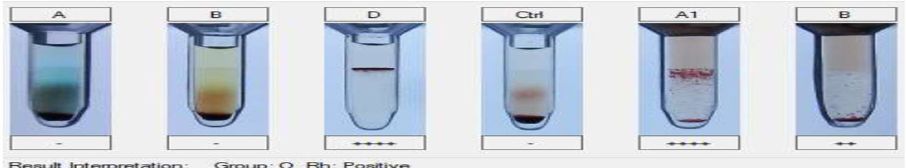 |
Figure 10: G (4+): Rh, A-serum; G (2+): B-Serum; Negative (-): A-cells, B-cells, Ctrl |
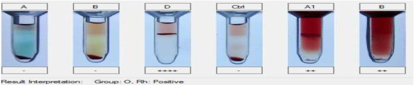 |
Figure 11: G (4+): Rh; G (2+): A-serum, B-Serum; Negative (-): A-cells, B-cells, Ctrl |
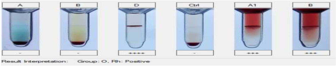 |
Figure 12: G (4+): Rh; G (3+): A-serum, B-Serum; Negative (-): A-cells, B-cells, Ctrl |
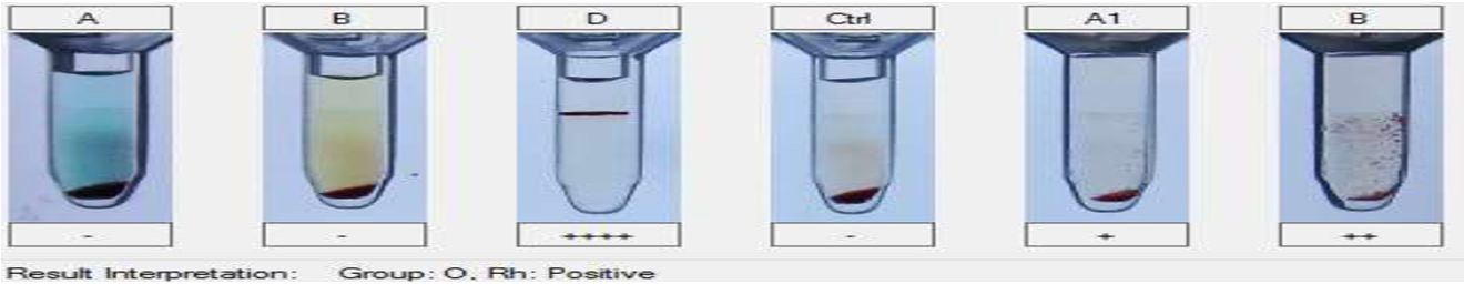 |
Figure 13:G (4+): Rh; G(2+): B-Serum; G(1+): A-serum; Negative(-): A-cells, B-cells, Ctrl |
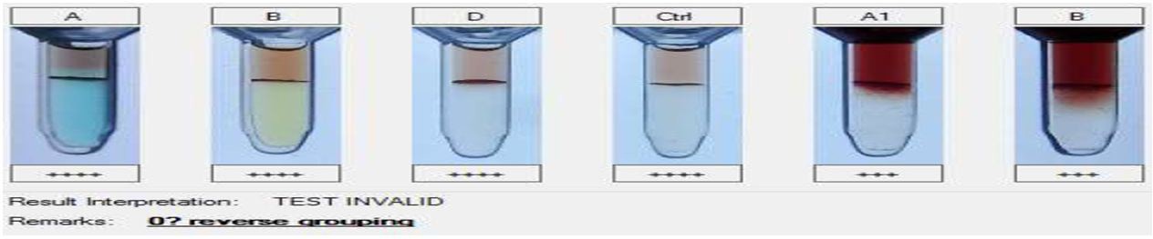 |
Figure 14:G (4+): A-cells, B-cells, Rh Ctrl; G (3+): A-serum, B-Serum |
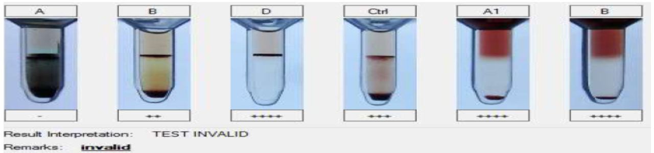 |
Figure 15: G (4+): Rh, A-serum, B-Serum; G (3+): Ctrl; G (2+): B-Cells; Negative(-): A-Cells |
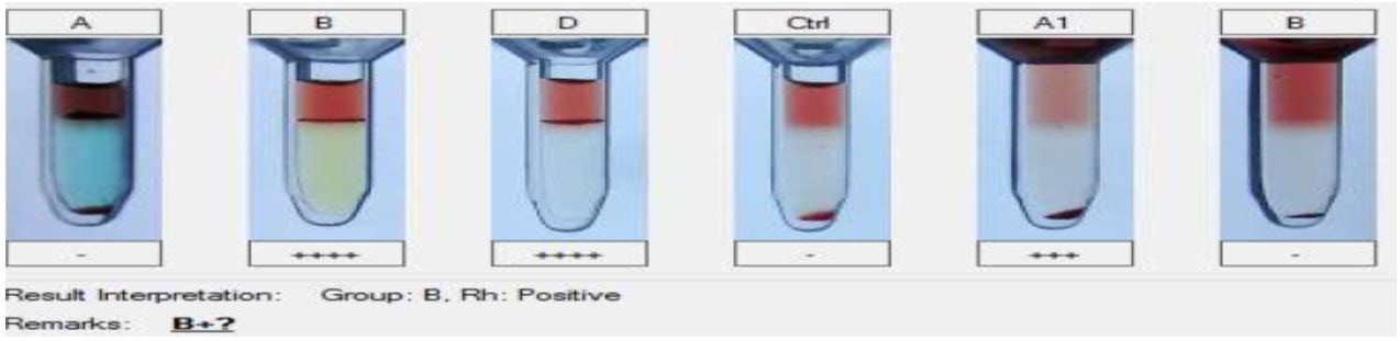 |
Figure 16: G (4+): B-Cells, Rh; G (3+): A Serum; Negative (-): A-Cells, Ctrl, B-serum |
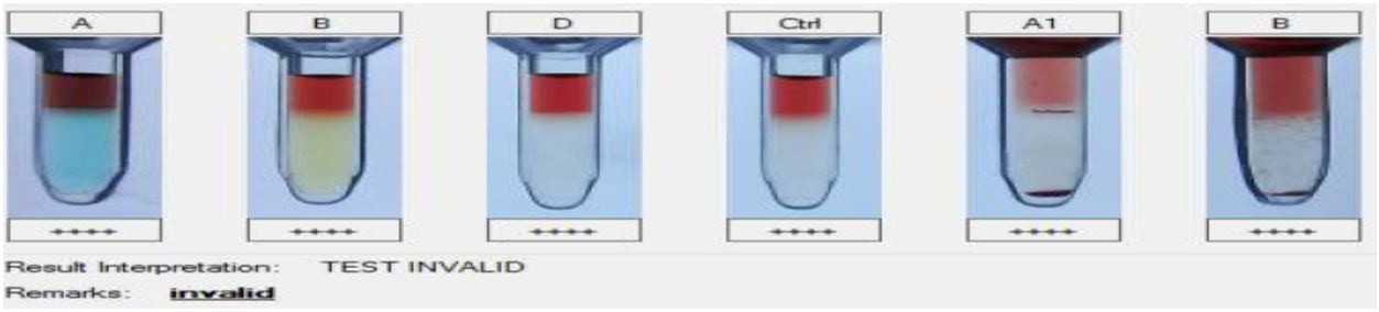 |
Figure 17:G (4+): A-Cells, B-Cells, Rh, Ctrl, A-serum, B-Serum |
Blood Group Type |
A |
B |
AB |
O |
Samples with correct results |
45 |
80 |
36 |
29 |
Table 1: Number of samples Showing blood group are as follows


