Open Access
Review Article
Max Screen >>
ISSN: 2348-9804
Copyright: © 2016 Manas B. This is an open-access article distributed under the terms of the Creative Commons Attribution License, which permits unrestricted use, distribution, and reproduction in any medium, provided the original author and source are credited.
Related article at Pubmed, Google Scholar
Bitemark analysis can be used for comparison of a known person’s dentition to a patterned injury which appears consisistent with a bitemark. This type of comparison is used to confirm or eliminate the identity of a suspect in relation to the bitemark. Because of the definite subjectivity in bite mark analysis it is considered as one of the most controversial of all forensic science investigations. There is a paucity of literature regarding the application and limitation of bite mark analysis in forensic science investigations. The aim of this paper is to review the current trends, applicability and guidelines of bite mark analysis.
Keywords: Bite marks; Forensic odontology; Bite mark analysis
Bite marks defined as, a physical alteration in a medium caused by the contact of teeth. A representative pattern left in an object or tissue by the dental structures of an animal or human. Bite marks occur primarily in sex-related crimes, child abuse cases, and offenses involving physical altercations, such as homicide [1]. Male victims are most often bitten on the arms and shoulders, while female victims are most commonly bitten on the breasts, arms and legs [2]. Figure 1
The current opinion is that bite mark can be useful in including or excluding possible suspects and ability to identify only one person as the biter. In mortal combat situations, such as the violence associated with life and death struggles between assailants and victims, the teeth are often used as a weapon [2,3].
These guidelines for bite mark analysis are result of a collective effort of the participants of the bitemark workshop of the American Board of Forensic Odontology assembled in Anaheim, California, February 18th through 20th, 1984. These guidelines are considered dynamic, not static, and will be modified as significant developments evolve [3]. Careful use of these guidelines in any bitemark analysis will enhance the quality of the investigation and conclusion [2].
In 1993, the ABFO Bitemark Workshop #2 Committee distributed a questionnaire on bitemark methodology. About half of their members responded to the Bitemark Methodology survey reviewed at the three day bitemark Workshop in San Antonio on February 12, 1994. The methods used by those that responded to collect and analyze bitemark evidence were presented. This project is an update of the efforts begun in the 1984 Bitemark workshop. This set of guidelines is not intended to invalidate the document generated as a result of the 1984 workshop [4].
Bite mark comparison is fairly new, however, going back to the mid-1970s. Police investigators have always noticed that at some crime scenes, criminals seem to leave their bite impressions on food products, chewing gum, or more commonly on the skin of their victims, especially in cases of robbery, rape, child abuse, and homicide. Ian A, et al. in 2000 studied 101 cases and observed that more than one bitemark was present in 48% of all the bite cases studied [5]. Bitemarks were found on adults in 81.3% of the cases and on children under 18 years-of-age in 16.7% of cases. Bitemarks were associated with the following types of crimes: murder, including attempted murder (53.9%), rape (20.8%), sexual assault (9.7%), child abuse (9.7%), burglary (3.3%), and kidnapping (12.6%).
There are seven types of bite marks which can be classified in Table 1 by four degrees of impression.
Identification of a suspect by matching his dentition with a bite mark found on the victim of a c1ime rests on the theory that each person’s dentition is unique. In this respect, bite mark comparisons are based on the same principle as the identification of a deceased person. This theory is accepted by courts [4,3].
Primarily bite marks can be classified into cutaneous human bitemark and prototypical human bitemarks [1].
Cutaneous Human Bitemark: An injury in skin caused by contacting teeth (with or without the lips or tongue) which shows the representational pattern of the oral structures (Figure 2).
Prototypical Human Bitemark: A circular or oval (doughnut) (ring-shaped) patterned injury consisting of two opposing (facing) symmetrical, U-shaped arches separated at their bases by open spaces [1,2]. Following the periphery of the arches are a series of individual abrasions, contusions and/or lacerations reflecting the size, shape, arrangement and distribution of the class characteristics of the contacting surfaces of the human dentition, different variations of prototypical bitemarks have been described [6] (Table 2).
It should be recognized that often the Forensic Odontologist is not involved in the initial examination and collection of the bitemark evidence. This does not necessarily preclude the ability of the Forensic Odontologist to render a valid opinion [7]. The below listed methods are not meant to be an all-encompassing list of preservation methods; however, it does list those methods that are used by the diplomates of the ABFO. The use of other methods of documenting the Bitemark evidence should be in addition to these techniques [8].
Saliva swabbings of the bite site should be obtained whenever possible. Obviously, certain circumstances may preclude the collection of this evidence. If the region had been washed prior to the opportunity to swab this procedure would not be possible. If swabbing the area would damage or alter the pattern, it should either not be done or accomplished only after all other preservation methods have been employed [5]. It is acceptable to use either cotton tip applicators or cigarette paper to gather this evidence. Other appropriate mediums may be used to collect this information. Control swabbings should be taken from other regions or port [9].
The bite site should be photographed using conventional photography and following the guidelines as described in the ABFO Bitemark Analysis Guidelines [2-4]. The actual photographic procedures should be performed by the forensic dentist or under the odontologist’s direction to insure accurate and complete documentation of the bite site. Color print or slide film and black and white film should be used whenever possible. Color or specialty filters may be used to record the bite site in addition to unfiltered photographs. Alternative methods of illumination may be used; Video/digital imaging may be used in addition to conventional photography. Off angle lighting using a point flash is the most common form of lighting and should be utilized whenever possible [10]. A light source perpendicular to the bite site can be utilized in addition to off angle lighting; however, care should be taken to prevent light reflection from obliterating mark details in photograph due to “wash out” due to light reflection. A light source parallel to the bite site can be utilized in addition to off angle lighting. A ring flash, natural light and/or overhead diffuse lighting can be utilized to off angle lighting. An ABFO No. 2 scale should be utilized whenever possible. The placement of the scale should follow the guidelines as established in the ABFO Bitemark Analysis Guidelines [8,11].
Impressions of the bite site should be taken when indicated according to the ABFO Bitemark Analysis Guidelines [9]. A backing material should be used to maintain the contour of the impression site. When the bite site is accessible to the victim’s dentition impressions of the victim’s teeth should be obtained. It may be useful if victim had bitten the assailant [12].
The bite site should be preserved when indicated following proper stabilization prior to removal. The resection of the tissue should follow all other evidence collecting procedures. 10% formalin is the common fixative used [3,6,12].
Both in the case of a living victim or deceased individual, the odontologist should determine and record certain vital information [8,13] (Table 3).
Several methods of bite marks comparison have been documented in the literature. All the documented methods are chiefly comprised of three steps [14].
• Registration of bite marks and suspect’s dentition.
• Comparison of the bite mark and dentition.
• Analysis of similarity and dissimilarity.
Registration of the bite mark by photography is used in all cases; the photographs are then enlarged to life-size proportion for comparison. However, ‘’a potential bite must be recognized early, as the clarity and shape of the mark may change in a relatively short time in both living and dead victims, where bite indentations (three-dimensional bite marks) are present in the skin tissue, impressions may be obtained; these are used to reproduce models of the bitemark, which can then be used for comparison [6,9,15]. An acetate overlay is an outline of the biting edge of someone’s teeth as traced onto and seen on a clear transparency (Figure 3).
Recovery of salivary DNA has been the main focus of biological techniques in bitemark analysis. The advantage of this method is that the DNA recovered from the saliva on the bitemark is usually sufficient to produce a profile. There are certain areas of concern. Extreme environmental circumstances are to be taken into consideration. The salivary DNA may be highly degraded. It can be assaulted by the environment. A new area in biological techniques is the use of the bacterial fingerprint [2-4,7,9,11]. There are over 2000 recorded species of oral bacteria and each individual has a unique bacterial population [16]. A bacterial fingerprint or a bacterial profile can be generated by recording the different species of bacteria present. This can be used to create a database in future. As of now this technique can be used to match a suspect’s bacterial profile. This technique is still nascent and has not undergone a lot of active research [9,17].
Skin is a very resilient and elastic material. The skin stretches during the bite due to elastic fibres in the dermis. This effect is only temporary. Due to damage control action taken by the skin cells, the skin reverts back to its normal position if it is not affected beyond its threshold limit. It also has the capacity to form a new layer of skin on the affected area if it is affected beyond its threshold [3]. Biting dynamics lead to different appearances of the bitemarks created by the same biter in cases involving multiple bitemarks [2,7]. There is definite subjectivity in bitemark analysis. This interpretative property of the science has led to questions about the validity, accuracy and reliability of bitemark analysis [3].
Forensic odontology and Molecular Biology has great importance to the expert practice when we think of a dentist enters in forensic investigation team for a bite mark case. It is necessary to broaden the pertinent studies of the theme, in order to establish protocols to allow additional tools in criminal investigation. Bite mark analysis has been approved by court but still, it has a long way to go before it becomes a mainstream form of expert evidence.

![]()

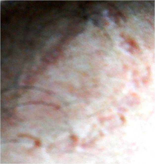 |
Figure 1: Bite marks on right arm of a rape victim (Courtesy Bhaskaran V University of Sri Jayewerdenepura Sri Lanka) |
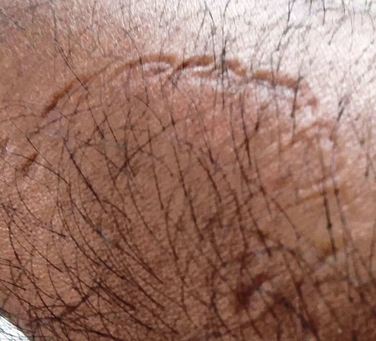 |
Figure 2: A cutaneous human bitemark reveals a representational pattern of the dentition. (Courtesy Bhaskaran V University of Sri Jayewerdenapura Sri Lanka) |
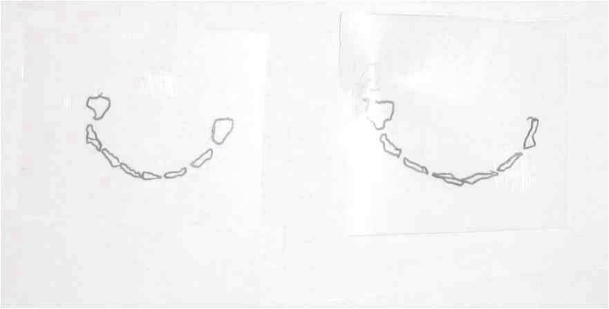 |
Figure 3: Bite marks registration transferred on transparency |
Types |
|---|
1. Hemorrhage -- small bleeding spot |
2. Abrasion -- undamaging mark on skin |
3. Contusion -- ruptured blood vessel, bruise |
4. Laceration -- punctured or torn skin |
5. Incision -- neat puncture of skin |
6. Avulsion -- removal of skin |
7. Artifact -- bitten-off piece of body |
Degrees of impression |
|---|
1. Clearly defined -- significant pressure |
2. Obviously defined -- first-degree pressure |
3. Quite noticeable -- violent pressure |
4. Lacerated -- skin violently torn from body |
Table 1: Types of bite marks and degrees of impression |
Patterns |
Features |
Central echymosis |
Discoloration of the skin of the bitten area. The reasons may be: |
Linear Abrasions, Contusions or Striations |
Represent marks made by either slipping of teeth against skin or by imprinting of the lingual surfaces of teeth. The term drag marks is in common usage to describe the movement between the teeth and the skin while lingual markings is an appropriate term when the anatomy of the lingual surfaces are identified. Other acceptable descriptive terms include radial or sunburst pattern. |
Double Bite |
A "bite within a bite" occurs when skin slips after an initial contact of the teeth and then the teeth contact again a second time. |
Peripheral Ecchymosis |
Due to excessive, confluent bruising. |
Partial bitemarks |
May represents 3 distinctive patterns |
Indistinct/Faded Bitemarks |
May shows different characteristic patterns. • Fused Arches - collective pressure of teeth leaves arched rings without showing individual tooth marks. • Solid - Ring pattern is not apparent because erythema or contusion fills the entire center leaving a filled, discolored, circular mark. • Closed Arches - the maxillary and mandibular arch are not separate but joined at their edges. • Latent – Can be visualized only with special imaging techniques. |
Superimposed or Multiple Bites. |
This pattern may be a result of multiple bite marks at the same site. |
Table 2: Different types of prototypical bite patterns |
|
Demographics • Name of victim • Case Number • Date of examination • Referring agency • Person to contact • Age of victim • Race of victim • Sex of victim • Name of examiner(s) |
To be filled by Forensic odontologist. |
Location of Bitemark • Describe anatomical location • Describe surface contour: flat, curved or irregular • Describe tissue characteristics A. Underlying structure: bone, cartilage, muscle, fat B. Skin: relatively fixed or mobile |
|
Shape • The shape of the bitemark should be described; e.g. essentially round, ovoid, crescent, irregular, etc. |
|
Color • The color should be noted; e.g. red, purple, etc. |
|
Size • Vertical and horizontal dimensions of the bitemark should be noted, preferably in the metric system. |
|
Type of Injury • Petechial hemorrhage • Contusion (ecchymosis) • Abrasion • Laceration • Incision • Avulsion Signature of Odontologist |
|
Table 3: Bite mark analysis guidelines |
|


