Open Access
research Article
Max Screen >>
ISSN: 2454-3276
Copyright: © 2015 El Meligy OA. This is an open-access article distributed under the terms of the Creative Commons Attribution License, which permits unrestricted use, distribution, and reproduction in any medium, provided the original author and source are credited.
Related article at Pubmed, Google Scholar
Aim: To evaluate microleakage of pit and fissure sealant after using four different pit and fissure preparation techniques.
Materials and Methods: Eighty exfoliated primary molars with no clinical evidence of caries were randomly divided into 4 groups of 20 each. Teeth were prepared using 1 of 4 occlusal surface treatment prior to placement of Conseal F opaque light cured sealant. The teeth were thermocycled between 5 ± 2 oC and 55 ± 2 oC, for 500 cycles with dwell time of 30 seconds then stored in 0.9% normal saline. All teeth were sealed apically and coated within 1.5 mm of the sealant margin with 2 layers of nail varnish. The teeth were immersed in a 1% solution of methylene blue for 24 hours to allow dye penetration. Three buccolingual cuts parallel to the long axis of the teeth were made yielding 4 sections and 6 surfaces per tooth for analysis. The surfaces were scored 0 to 3 for extent of microleakage using a binocular microscope at 25x magnification.
Results: There was no significant difference in microleakage between the four groups although the air abrasion and acid etching exhibited the least microleakage followed by enameloplasty and acid etching then the self etching adhesive system, and the highest microleakage was pumice and acid etching.
Conclusions: Neither air abrasion and enameloplasty followed by acid etching nor the self etching adhesive system produced significantly less microleakage than the traditional pumice prophylaxis with acid etching technique.
Keywords: Pumice; Enameloplasty; Dentin Adhesive; Air Abrasion; Microleakage
By (1995), Siegel [1] stated that sealants are an important dental caries prevention technology, ideally used in combination with patient education, effective personal oral hygiene, fluorides, regular dental visits. There is a growing need to seal primary molars, as demonstrated by Head Start's report that found that, for those children who had dental caries, 86% had caries of the pits and fissures of the molars [2].
The ability of a sealant to prevent microleakage is important because microleakage may support caries below the sealant. Pits and fissures that are successfully sealed may prevent or arrest early developing occlusal lesions [3]. Salivary pellicles, organic debris and handpiece lubricant oil have all been as potential contaminants of the tooth surface that may lead to marginal leakage or loss of the sealant [4]. Various pretreatment methods have been investigated with the intent to enhance the effectiveness of sealing the enamel surface [5].
Pumice prophylaxis before sealant application has been recommended in the past but opinion on this has lately shifted. Burrow 1990 [6] has shown that cleaning the fissures with flour of pumice using rotary instruments in a slow-speed handpiece followed by etching does not remove all of the pellicle and debris.
Alternative methods such as bur preparation and air abrasion have been proposed to better clean pits and fissures of debris. Enameloplasty or widening the fissures with rotary instrumentation permits better diagnosis of underlying decalcifications and improves sealant retention [7]. Air-abrasion was introduced in the 1950s, it uses a high-speed stream of purified aluminium oxide particles propelled by air pressure onto tooth surface. Stains and organic plugs found in most pits and fissures are removed. Enamel is roughened in a conservative and time-efficient manner which aids in sealant bonding [8]. However, marginal leakage studies have shown that air abrasion alone is not as effective as air abrasion coupled with acid etching in preventing microleakage [7,9].
More recently, the addition of a bonding agent to the traditional sealant technique has shown promising results as means of improving retention and microleakage. Hebling and Feigal [10] demonstrated a significant reduction in microleakage when singlebottle bonding agent was applied between a sealant and enamel interface. Self etching adhesive systems have also been developed which may be able to improve and simplify the sealant process further, thus warranting more investigation [11].
Researches have used dye penetration to evaluate potential sealant leakage and the susceptibility of a tooth to caries [12]. Conflicting results have been reported by studies comparing microleakage of sealants prepared by different methods [13-15]. Therefore, the search continues for the most effective enamel surface preparation to enhance sealant integrity. The null hypothesis tested was that there would be no difference in the microleakage of pit and fissure sealant after using four different pit and fissure preparation techniques on exfoliated primary molars with no clinical evidence of caries.
The aim of this invitro study was to compare the microleakage of pit and fissure sealant after using four different preparation
techniques:
1- Traditional pumice prophylaxis and acid etching.
2- Fissure enameloplasty and acid etching.
3- Self-etching adhesive system.
4- Air abrasion and acid etching.
Eighty carious-free and unrestored exfoliated primary molars were stored and refrigerated in 0.1% chloramine. They were randomly divided into 4 groups of 20 each.
Group I: Traditional pumice prophylaxis and acid etching. All fissures were cleaned for 15 seconds with an aqueous slurry of 5g pumice/4ml water using a disposable rotating bristle brush in a slow-speed, contra-angle handpiece. The teeth were rinsed, dried and acid etched with 37% phosphoric acid for 60 seconds as recommended by the sealant manufacturer.
Group II: Enameloplasty and acid etching. The pits and fissures were opened with a ¼-round bur in a low speed handpiece to an approximate diameter of the bur and then acid etched.
Group III: Self-etching primer adhesive group (One-Up Bond F Plus, Tokuyama Dental Corp. 38-9, Taitoul-chome, Tokyo, Japan). One drop of bonding agent A was mixed with bonding agent B in a mixing well, mix thoroughly and evenly until the mixed bonding agents turns pink. Apply the mixed bonding agent on enamel, wait for 20 seconds, light cure for 10 seconds.
Group IV: Air abrasion and acid etching. The KCP air abrasion unit (American Dental Technologies Troy, MI 48084 USA) was used according to manufacturer's directions with 27.5um aluminium oxide particles at 120 psi. The nozzle tip of approximately 0.375 mm diameter was held 3 to 4 millimeter from the target site. The kinetic stream was applied for 5 to 10 seconds. At the end, frosted finished surface was obtained and then acid etched.
An opaque light cured sealant (Conseal F/SDI Victoria, Australia) was applied to the occlusal pits and fissures of all teeth in accordance with the manufacturer's instructions. The sealant was cured for 15 seconds according to the recommendations of the sealant manufacturer. Each sealant was checked with an explorer for complete coverage and retention. All sealed teeth were stored in 0.9% normal saline less than 24 hours before thermocycling.
All of the teeth were thermocycled between 5 ± 2 oC and 55 ± 2 oC for 500 cycles with a dwell time of 30 seconds then stored in 0.9% normal saline [16]. The teeth were dried and the apices sealed with sticky wax. All tooth surfaces were painted within 1.5mm of the sealant margin with a layer of nail varnish and allowed to dry. A second coat of nail varnish was applied and allowed to dry. The teeth were immersed in a 1% methylene blue solution for 24 hours at 37 oC to allow dye penetration into possible gaps between enamel and sealant.
Upon removal from the dye, the teeth were rinsed with distilled water, positioned, and secured on glass slabs using sticky wax prior to sectioning. Three buccolingual sectioning cuts parallel to the long axis of each tooth were made, yielding 4 sections and 6 surfaces per tooth for analysis. A water-cooled 0.3mm thick diamond watering blade mounted on a high-speed saw (Isomet 1000; Buehler, Lake Bluff, IL, USA) was used. The depth of dye penetration was evaluated by a single examiner blinded to the treatment regimen [17] using a binocular microscope at 25x magnification according to the method described by Overbo and Raadol [18]. The scoring method was (Figure 1):
Score 0 = no dye penetration.
Score 1 = dye penetration restricted to the outer half of the sealant.
Score 2 = dye penetration to the inner half of the sealant.
Score 3 = dye penetration into the underlying fissure.
Each surface score was determined by the greatest dye penetration detected on the buccal occlusal and/or lingual occlusal fissure wall. The overall score for each tooth equaled the highest score of the 6 surfaces. Mean microleakage scores and standard errors were calculated for each treatment group.
The Kruskal-Wallis test was used to statistically analyze for significant differences in microleakage between treatment groups. The Ethical Committee at the Faculty of Dentistry, Alexandria University approved the research protocol.
Table 1 shows the microleakage scores according to section level analysis with 6 surface measurement per tooth totaling 480 scores. Statistical analysis using Kruskal-Wallis test indicated no significant difference between the pumice, enameloplasty, dentine adhesive and air abrasion methods. No leakage (score 0) was found on 361 of 480 surfaces (75.2%). Dye penetration restricted to the outer half of the sealant (score 1) was noted in 74 of 480 views (15.4%).
Thus, about 90% of the sealant sections had minimal to no leakage. Dye penetration to the inner half of the sealant (score 2) was noted in 35 of 480 views (7.3%). Dye penetration into underlying fissure (score 3) was noted in 10 of 480 views (2.1%). The depth of dye penetration is shown in Figures 2, 3, 4 and 5.
The diamond blade was of sufficient thickness (0.3mm) to prevent adjacent surfaces from being "mirror images" of each other. For example, there were many instances in which one surface displayed microleakage, and the adjacent sectioned surface had no microleakage.
According to Kruskal-Wallis test there was no statistically significant difference (p > 0.05) between the 4 treatment groups represented by the mean microleakage scores in Table 2. However, the least mean of microleakage scores was Group IV (Air abrasion acid etching) (0.29 ± 0.6), followed by Group II (Enameloplasty and acid etching) (0.33 ± 0.65) and group III (self etching dentine adhesive) (0.35 ± 0.69). The highest mean of microleakage scores was Group I (Pumice and acid etching) (0.48 ± 0.86).
The null hypothesis was not rejected, since there was no significant difference in microleakage of pit and fissure sealants after using four different preparation techniques.
A variety of fissure preparation methods have been used prior to sealant placement in an attempt to successfully inhibit microleakage and maximize retention. While sealants have gained acceptance as their retentive and microleakage properties have improved, their clinical success rate still remains less than ideal. A 5% to 10% annual sealant failure rate was reported following analysis of multiple studies [19]. Consequently, the search for alternative preparation method has continued to be an ongoing challenge.
In this study, eighty exfoliated primary molars with no clinical evidence of caries were randomly divided into 4 groups based on the surface pre-treatment technique used. An opaque light cured sealant was applied according to the manufacturer's instructions. The sealed teeth were thermocycled between 5 ± 2 oC and 55 ± 2 oC, for 500 cycles with dwell time of 30 seconds [16], then immersed in a 1% solution of methylene blue. The dye leakage method was used in this study because it is simple, inexpensive and do not require the use of complex laboratory equipment [20].
The findings of this study has some implications regarding sealant placement. First, it suggests that dentists may be using more aggressive sealant preparation technique on sound, healthy tooth structure than is necessary. Secondly, air abrasion is not without disadvantages, as it often creates a substantial dust powder in the work area that requires additional time to clean up.
In the present study, no significant difference in sealant microleakage was found between pumice, fissure enameloplasty, self etching adhesive system and air abrasion. This study results are supported by those of Mentes and Gencoglu [12] who used the same scoring system by Overbo and Raadol [18] to analyze dye penetration. Moreover, the air abrasion/etch group produced the lowest microleakage scores in both studies but not significantly lower than the other 3 treatment methods.
Similar results have been obtained in other microleakage studies. Xalabarde et al [14] demonstrated no significant difference in microleakage when comparing enameloplasty and etch to pumice and etch. They evaluated 2 different types of burs, the soreness diamond and ¼-round carbide, but neither produced significantly marginal leakage. In a study by Lupi-Pegurier et al [7], the extent of microleakage was comparable between bur and air abrasion preparation methods when acid etch was employed. A study by Guirguis et al [9] yielded similar results. The marginal leakage of preventive resin restorations placed in teeth prepared with air abrasion alone was compared to resin restorations placed after bur preparation or air abrasion coupled with acid etching. Significant microleakage was observed in the non-etched, air abraded specimens, while the resin prepared by bur/etch or air abrasion/etch produced similar microleakage results that were significantly less than air abrasion alone. Also, Sancakli et al [17] recorded that air abrasion followed by acid etching, as well as conventional acid etching, provided a sufficient seal.
In contrast, Hatibovic-Kofman et al [13] reported bur preparation coupled with acid etching to be significantly better at reducing microleakage than pumice and etch. The difference in findings may be due to the use of different burs and preparation depths. Geiger et al [21] demonstrated that the deeper the level of sealant penetration, the lower the probability of microleakage.
A variety of fissure preparation methods have been used prior to sealant placement in an attempt to successfully inhibit microleakage and maximize retention. While sealants have gained acceptance as their retentive and microleakage properties have improved, their clinical success rate still remains less than ideal. A 5% to 10% annual sealant failure rate was reported following analysis of multiple studies [19]. Consequently, the search for alternative preparation method has continued to be an ongoing challenge. In this study, eighty exfoliated primary molars with no clinical evidence of caries were randomly divided into 4 groups based on the surface pre-treatment technique used. An opaque light cured sealant was applied according to the manufacturer's instructions. The sealed teeth were thermocycled between 5 ± 2 oC and 55 ± 2 oC, for 500 cycles with dwell time of 30 seconds [16], then immersed in a 1% solution of methylene blue. The dye leakage method was used in this study because it is simple, inexpensive and do not require the use of complex laboratory equipment [20].
The findings of this study has some implications regarding sealant placement. First, it suggests that dentists may be using more aggressive sealant preparation technique on sound, healthy tooth structure than is necessary. Secondly, air abrasion is not without disadvantages, as it often creates a substantial dust powder in the work area that requires additional time to clean up. Lending support to this study's finding is an in vitro sealant study by Brown and Barkmeier [22] that found no significant difference in bond strength between air abrasion and acid etch compared to acid etch alone. Similarly, a retention study conducted on extracted molars found that all acid-etched groups, which included a pumice, air abrasion, hydrogen peroxide and acid-etch alone group generated equivalent mean shear bond strengths [23].
In contrast, Geiger et al [21] demonstrated on extracted molars that sealant retention was significantly improved by bur preparation compared to non-prepared fissures. Bagherian et al [24] concluded that the use of fissurotomy bur and pumice prophylaxis accompanied with acid etching appears to have a more successful reduction of microleakage than acid etch alone.
Circumstances do present where bur or air abrasion preparation is obviously warranted. Suspected carious fissures should be prepared or explored by enameloplasty or air abrasion prior to sealant application to disclose potential caries. However, several studies have provided evidence that inadvertent sealing over carious lesions leads to arrest and not to caries progression [25-27]. The need to routinely air abrade or mechanically prepare fissures prior to sealing is not supported in the literature.
This study's results suggest that the method of pit and fissure cleaning is not the critical step in the sealant technique. It is more likely conditions such as both isolation and moisture control, eruption status of tooth, patient co-operation and a proper etch that determine sealant success.
This study did not omit acid etching in the air abrasion group because the majority of previous studies have consistently demonstrated that the roughened surface produced by air abrasion alone lacks the seal obtained with acid etching [5,9,12,16]. The literature appears to support acid etching as the treatment of choice over air abrasion alone.
The increased enamel resin tag formation that has been observed in SEM studies is one of the main benefits of acid etching [28,29]. Resin tag formation provides micromechanical retention of the sealant to enamel interface and serves as the primary mechanism by which sealants bond to enamel fissures [30]. This study's findings suggest that it makes no difference what type of fissure preparation is used as long as it is followed by acid etching prior to sealant placement. More recently, the addition of a bonding agent to the traditional sealant technique has shown promising results as a mean of improving retention and microleakage. Feigel et al [31] found single-bottle bonding agents to reduce the usual risk of retention failures of occlusal sealants. Another study demonstrated a significant reduction in microleakage when a dentin-bonding agent was applied between a sealant and salivacontaminated enamel interface [10]. However, these studies used adhesive resins that were placed following conventional acid etching, and the products themselves were not acidic resins. In the present study, it was anticipated that the use of an acidic resin primer would provide significantly higher protection against marginal microleakage as that seen with conventional acid-etching techniques. However, the results did not prove this assumption true.
A study by Perry and Rueggeberg [32] yielded similar results. The microleakage patterns observed when the acidic resin primer was used, are more likely the result of inability to seal the margin with a well-cured resin than by supplying an adequately etched enamel surface. Lack of marginal seal with the self-etching primer adhesive system (Prompt-L-Pop) used on extracted human teeth was also noted by Pradelle-Plasse et al [33]. However, in that study, leakage of Class V restorations was evaluated on an abraded enamel surface, unlike the present study where untreated occlusal surface were tested.
This investigation was performed in vitro using exfoliated primary molars. A controlled clinical trial is needed to further study the 4 preparation methods. Until microleakage and retention studies demonstrate conclusively one preparation method to be more effective than another at enhancing sealant success, it would seem prudent to continue using the traditional pumice and acid-etch technique. Based on the present findings, routine removal of healthy sound tooth structure prior to sealant placement does not seem justified.
Neither air abrasion and enameloplasty followed by acid etching nor the self etching adhesive system produced significantly less microleakage than the traditional pumice prophylaxis with acid etching technique. This invitro comparative study suggests that the conventional pumice prophylaxis with acid etching technique should remain the standard of practice for cleaning fissures prior to sealant placement.

![]()

 |
| Figure 1: Schematic diagram for dye penetration scoring as described in text |
 |
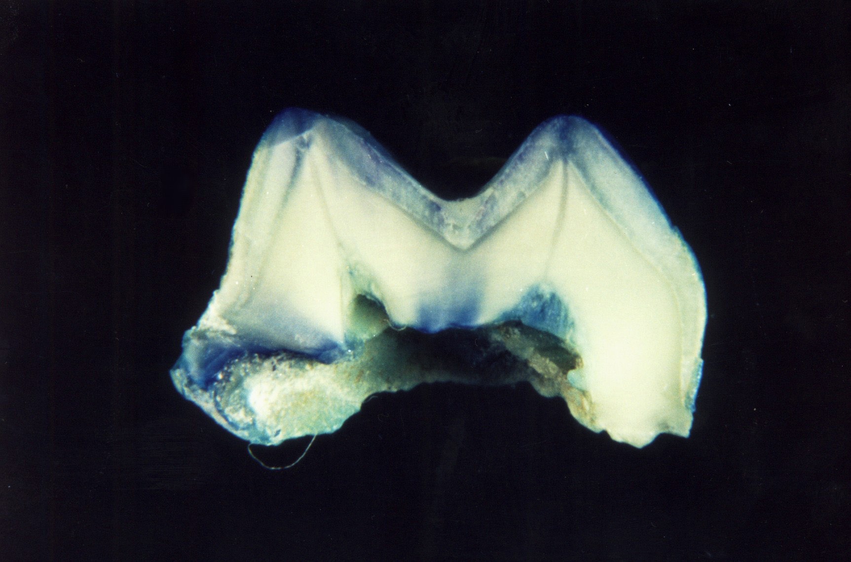
| Figure 2: Shows no dye penetration (score 0) |
 |
| Figure 3: Shows dye penetration restricted to the outer half of the sealant (score 1) |
 |
| Figure 4: Shows dye penetration to the inner half of the sealant (score 2) |
 |
| Figure 5: Shows dye penetration into underlying fissure (score 3) |
| Dye penetration | |||||
|---|---|---|---|---|---|
| Preparation Method | 0 | 1 | 2 | 3 | Total |
| Pumice | 87 | 14 | 14 | 5 | 120 |
| Enameloplasty | 91 | 19 | 9 | 1 | 120 |
| Dentine adhesive | 90 | 21 | 6 | 3 | 120 |
| Air abrasion | 93 | 20 | 6 | 1 | 120 |
| Total | 361 | 74 | 35 | 10 | 480 |
| 0 = no dye penetration. 1 = dye penetration restricted to the outer half of the sealant. 2 = dye penetration to the inner half of the sealant. 3 = dye penetration into the underlying fissure. Table 1: Distribution of section-level microleakage scores. |
|||||
| Preparation Method | N | Mean | SD | Std. Error |
|---|---|---|---|---|
| Pumice | 120 | 0.48 | 0.86 | 7.8E-02 |
| Enameloplasty | 120 | 0.33 | 0.65 | 5.96E-02 |
| Dentine adhesive | 120 | 0.35 | 0.69 | 6.33E-02 |
| Air abrasion | 120 | 0.29 | 0.60 | 5.4E-02 |
| Total | 480 | 0.36 | 0.71 | 3.24E-02 |
| The median equals 0 with all four different pit and fissure preparation methods. Table 2: Mean microleakage scores-section level analysis |
||||


