Open Access
Case report
Max Screen >>
ISSN: 2348-9820
Copyright: © 2019 Silva IV. This is an open-access article distributed under the terms of the Creative Commons Attribution License, which permits unrestricted use, distribution, and reproduction in any medium, provided the original author and source are credited.
Related article at Pubmed, Google Scholar
Ameloblastoma is a benign odontogenic tumor, comprising 1% of all tumors and cysts of the jaws. A lot of the patients are asymptomatic and most cases are diagnosed when the swelling is already evident. It is a locally aggressive tumor with a high recurrence rate when conservative surgical treatment is applied (up to 80%). That is why current treatment consists of wide resection of the lesion with safety margins of healthy bone beyond radiographic limits.
After tumor excision, it is important to reconstruct the mandibular defect, in order to restore both function as well as aesthetics of the jaw. Free vascularized bone flaps are widely recognized as the optimal reconstruction. Ideally, the flap must provide enough bone for reconstruction of the defect and to accommodate osseointegrated dental implants. Fibula and iliac crest are the most commonly used free flaps for mandibular reconstruction.
The major advantages of the iliac crest flap are its natural curvature, the abundance of vertical and horizontal height as well as quality of the bone and the inconspicuous donor site scar. Although this flap dissection is considered to be more difficult, this depends a lot on the surgeon experience.
Currently, the mechanisms involved in the occurrence of bilateral parotid tumors are still unclear and its development may be coincidental.
We present two cases of mandible ameloblastoma, one with 6 months of follow-up and the other with 4 years of follow-up, that were successfully managed with hemimandibulectomy and immediate reconstruction using free vascularized iliac crest flap.
Keywords:Ameloblastoma; Mandibular Reconstruction; Iliac Crest Free Flap; Microsurgery
List of abbreviations: CT: Computed Tomography
Ameloblastoma is a benign odontogenic tumor of the jaws, arising from remnants of dental lamina [1], characterized by the tendency for local recurrence if not adequately removed [1,2]. It represents about 1% of all tumors and cyst of the jaws [2,3] and is the second most common odontogenic tumor [1]. The preferred location is the mandible (about 80%) [1], especially in the posterior region [1,4].
The diagnosis is often delayed probably due to its painless slow-growing character [2]. Depending on how early the patient presents for evaluation, the clinical manifestation may range from an innocuous intraoral swelling that the patient is unaware of to a grotesque facial swelling [5]. With increasing size, other complications may arise, including loosening of teeth, malocclusion, paresthesia, pain, soft tissue invasion, facial deformity, reduced mouth opening, difficulty with mastication, and airway obstruction [1].
Radiographically, ameloblastoma typically presents as a corticated multilocular radiolucency with a so-called soap-bubble or honeycomb pattern and a unilocular appearance is less common [1]. Computed Tomography (CT) is the gold standard for evaluation of ameloblastomas, being a helpful tool in treatment planning [5].
Current treatment is wide surgical excision including an area of bone beyond radiographical margins [1]. Conservative surgery (such as enucleation and curettage) is associated with high recurrence rates (21-80%) [1,2].
After tumor excision, it is important to reconstruct the mandibular defect, in order to restore both function and aesthetics [6,7]. Starting from the 1980s, the development of microvascular techniques has dramatically changed the prospective for reconstruction of complex defects of the mandible [7]. Free vascularized bone flaps are widely accepted as the optimal reconstruction, providing the best restoration of oral function, cosmetic contour and quality of life and have become the standard of care [6].
The aim of the present article is to report two cases of mandible ameloblastoma, one with 6 months of follow-up and the other with 4 years of follow-up, with emphasis on the surgical treatment and reconstruction of the mandibular defect with a free vascularized iliac crest flap.
A 42-year-old African woman was referred to the Plastic Reconstructive and Craniomaxillofacial Surgery and Microsurgery Unit of Centro Hospitalar of Vila Nova de Gaia/Espinho, with a three-year history of discomfort and gradually increasing swelling on the left side of the mandible.
A panoramic radiograph (Figure 1) showed an intraosseous multilocular radiolucent lesion of the left mandible, extending from the left molar region to the left condyle.
Clinical examination revealed discreet facial asymmetry due to swelling on the left mandibular region. An intraoral evaluation showed a swelling on the posterior left mandible that was hard and not tender on palpation and was covered by intact mucosa.
Incisional biopsy was performed under local anesthesia and histopathologic study confirmed the suspected diagnosis of ameloblastoma.
CT of the jaws (Figure 2) revealed a well-defined multilocular expansile mandibular lesion, extending from the first molar region to the left mandibular ramus and condyle.
Under general anesthesia, a left hemimandibulectomy was performed from the 3.4 tooth and including the condyle, along with immediate reconstruction using left iliac crest vascularized free flap. The left facial artery and left external jugular vein were chosen as the recipient vessels. The osseous flap was fixated with a titanium reconstruction plate and screws.
Histopathologic examination of the resected mandibular segment confirmed the diagnosis of ameloblastoma.
The postoperative period was uneventful, and the patient was discharged from the hospital after 18 days.
The patient underwent oral rehabilitation with two endosseous dental implants, three years after the surgery.
Periodic clinical and imagiological follow-up was performed (Figure 3).
The last imagiologic follow-up (Figure 4) was performed at 3.5 years post-surgery, revealing complete integration of the flap and no evidence of tumor recurrence.
At the time of this report, four years post-surgery, the patient was pleased with the aesthetic and functional result, with acceptable facial symmetry and jaw movement (Figure 5). There were no signs of tumor recurrence.
A 27-year-old Caucasian man reported with a gradually growing painless swelling on the left mandibular ramus. Before he was referred to our Hospital, he had visited a private clinic and underwent an incisional biopsy of the mandibular lesion, which yielded the diagnosis of ameloblastoma.
On clinical examination, the patient presented discreet facial asymmetry and intraoral inspection revealed a solitary swelling on the left lower posterior vestibule, extending from the retromolar region to the posterior border of the mandibular ramus. On palpation, the swelling had hard consistency and there was no tenderness.
CT (Figure 6) revealed a well-defined osteolytic expansile lesion in the left mandibular ramus and angle with unilocular appearance, causing expansion of the mandibular ramus, and the unerupted third molar was present within the radiolucency
Under general anesthesia, a left hemimandibulectomy was performed from the tooth 3.5 including condyle. The reconstructive procedure was executed on the same surgical setting. An ipsilateral osseous iliac crest flap was harvested. Preoperative virtual 3D biomodelling techniques where applied in order to plan the exact dimensions, angles and contour of the iliac crest flap. Left facial artery and left external jugular vein were used as recipient vessels and the osseous flap was fixated using a titanium reconstruction plate and screws (Figure 7).
Histopathologic examination of the resected mandibular segment confirmed the diagnosis of ameloblastoma.
There were no significant complications on the postoperative period, and the patient was discharged from the hospital after 16 days.
The last imagiologic follow-up (Figure 8) was performed at five months post-surgery, revealing integration of the flap and no evidence of tumor recurrence.
At the time of this report, six months post-surgery, it was evident that the reconstructive procedure gave adequate support to the soft tissue, with acceptable cosmetic result and jaw movement close to normal (Figure 9). There were no signs of tumor recurrence.
The next phase of treatment will be the oral rehabilitation with dental implants.
Although ameloblastoma is considered a benign tumor, it is locally aggressive and has a high recurrence rate if not adequately removed [2].
A lot of the patients are asymptomatic, and the early manifestation is as a slow-growing painless expansion. Therefore, most patients are diagnosed in the late period when the swelling is already evident, or, by chance, during routine radiographic exams [3].
The management of ameloblastoma is still controversial. The treatment of choice is surgical resection, however, no unanimous consensus exists concerning the extension of surgery. The surgical approach can be divided in conservative and radical methods. Conservative surgical procedures, including enucleation, curettage and marsupialization [3,5], preserve the patients’ normal tissues and minimizes facial disfiguration [5], however, they are prone to higher recurrence rates [2,5].
The radical surgical treatment involves en bloc tumor resection with wide safety margins [5] removing the surrounding tissues, in order to reduce the risk of leaving neoplasic cells behind, which would lead to recurrence [3].
As the face plays a central role in our daily interactions, a large mandibular defect means a significant loss in the quality of life, unless it is reconstructed successfully [8]. This radical modality of treatment implies an adequate immediate or delayed reconstruction of the surgical defect, in order to restore masticatory function and facial harmony [3]. With immediate reconstruction, there is less infection, scarring, contraction and morbidity [3].
Although there are many options for mandibular reconstruction (including reconstruction plates and nonvascularized bone grafts), vascularized free flaps provide much superior results in terms of the amount and durability of supplied bone [8]. Consequently, vascularized bone transfer has become the gold standard for mandibular reconstruction [8].
During the past decades, a variety of donor sites for vascular bone flaps and soft tissue have evolved. Ideally, the flap must provide adequate bone with sufficient length, thickness and width for reconstruction of the bone component of the defect and to accommodate osseointegrated dental implants.
Fibula and iliac crest are the most commonly used free flaps for mandibular reconstruction [3,8]. The main advantages of fibula flaps are quicker flap harvest with less blood loss (as the flap dissection is performed under tourniquet) [3,8], enough length of fibular bone segment for any length of mandibular defect [3,8], and a long pedicle (up to 8 cm) [3,8]. Disadvantages of fibula flaps are the straightness of the bone, requiring osteotomies for curvature, which is time consuming [3,8], and inadequate height of the fibular bone in comparison to a dentulous mandible, which can be a problem when dental implants are planned [3,8]. Because of its straight shape, fibula flap is more suitable for anterior mandibular defect [3]. Although the donor site functional morbidity is relatively mild, it is a significant aesthetic problem, particularly for woman [8].
The iliac crest is the other most frequently used donor site. The major advantages are its intrinsic curvature, which means that it is already precontoured for ipsilateral reconstruction [3,8], the abundance of vertical and horizontal height of bone available for mandibular contour and dental implant osseointegration [3,8] and the discreet donor site scar has a superior cosmetic result when compared with other choices [3,8]. Its major disadvantages are difficulty in dissection compared with fibular flap, with higher blood loss [3,8], presence of excess adipose tissue in obese patients, and risk of hernia [3]. Although this flap dissection is considered to be more difficult, this depends a lot on what type of procedure the surgeon is more accustomed to and can be overcome, in our opinion, with experience.
Donor site complications have been previously described as higher in iliac crest free flap than fibula, however, in our experience; there have been no significant differences.
In the cases described in this article, it was chosen a radical surgery treatment and hemimandibulectomy with resection of surgical margins beyond the radiographic margins of bone was performed. In both cases, the intrinsic curvature of the iliac crest was suitable for the hemimandibular defects that were created. In addition, as it was already mentioned, oral rehabilitation with osseointegrated dental implants is easier after reconstruction with iliac crest flap because of the abundant height of bone provided by this flap. There were no significant donor site complications observed in both patients.
The innovations around these case reports are more around the surgical planning of the reconstructions with custom-made surgical guides to use on the operation theatre. It is also true that iliac crest free flaps are not that commonly used in very large mandibular defects, where fibula flap normally are the option.
Although the mandibular condyle was resected in the reported cases, the function was not impaired, and the range of jaw opening was quite sufficient in both patients. In our experience, in large unilateral mandibular defects, a formal condyle reconstruction is not essential to obtain a good result and a satisfactory occlusion. We have patients with more than 15 years follow-up.
Oral rehabilitation is normally initiated around 6 months post-surgery, with radiological confirmation of good bone consolidation. Location of endosseous implants is based on CT-scans and, in some cases, with the use of stereolithography custom-made guides. Removal of the osteosynthesis hardware is sometimes deemed necessary for these procedures.
Ameloblastoma is an aggressive odontogenic tumor and current treatment consists of wide resection of the lesion with safety margins of healthy bone, as conservative surgery yields high recurrence rate.
Resection with simultaneous reconstruction will restore aesthetics as well as function.
Microsurgical reconstruction methods in addition to oral rehabilitation with osseointegrated dental implants is the current accepted treatment.
Both iliac and fibula free flaps should be considered for use in mandibular reconstruction.
Vascularized free iliac crest flap is a safe and reliable method to achieve functional and aesthetic mandibular reconstruction, particularly in lateral defects of the mandible.
Flap selection should be based on the advantages and disadvantages of each flap and also considering each patient individually, regarding age, gender, premorbid state, expectations and intention for oral rehabilitation.

![]()

 |
Figure 1: operative Panoramic Radiograph (case 1) – intraosseous multilocular radiolucent lesion of the left posterior mandible |
 |
Figure 2: Preoperative Computed Tomography (case 1) – radiolucent lesion in the left posterior mandible causing buccal and lingual cortical expansion |
 |
Figure 3: Postoperative Panoramic Radiograph, 1.5 years after surgery (case 1) – adequate symmetry and mandibular contour |
 |
Figure 4:Postoperative Computed Tomography, 3.5 years after surgery (case 1) (A) axial section; (B) 3D reconstruction, inferior aspect; (C) 3D reconstruction, frontal aspect |
 |
Figure 5: Follow-up photograph, 4 years after surgery (case 1) (A) frontal aspect; (B) side aspect; (C) adequate oral opening |
 |
Figure 6: Preoperative Computed Tomography (case 2) – unilocular radiolucent lesion in the left posterior mandible |
 |
Figure 7: Intraoperative photograph (case 2) - placement of iliac crest free flap fixated to the remaining mandible using a reconstruction plate and screws |
 |
Figure 8: Postoperative Computed Tomography, 5 months after surgery (case 2) (A) axial section; (B) 3D reconstruction, frontal aspect |
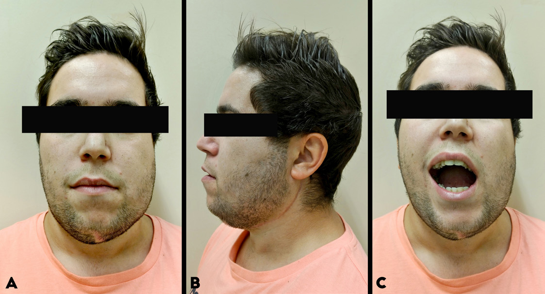 |
Figure 9: Follow-up photograph, 6 months after surgery (case 2) (A) frontal aspect; (B) side aspect; (C) adequate oral opening |


