Open Access
Case report
Max Screen >>
ISSN: 2348-9820
Copyright: © 2019 Horjeti E. This is an open-access article distributed under the terms of the Creative Commons Attribution License, which permits unrestricted use, distribution, and reproduction in any medium, provided the original author and source are credited.
Related article at Pubmed, Google Scholar
Stevens-Johnson syndrome (SJS) and toxic epidermal necrolysis (TEN) are closely-related, severe, acute life-threatening, drug-induced skin and mucosal disorders with a high mortality rate or long-term damages. These medical conditions are considered a delayed, type- IV hypersensitivity reaction and can be triggered by drugs, infections and malignancies.
SJS and TEN are considered to be two ends of the spectrum of one disease, accompanied by systemic symptoms, differing only by their extent of skin detachment. The classic form of these syndromes is featured by nonspecific prodromal symptoms and followed by cutaneous, mucosal and ocular damage, subsequently involving different systems such as: pulmonary, cardiovascular, renal and hepatic.
Since there is not a standardized guideline for the management of these disorders they require a multidisciplinary approach. Early diagnosis, recognition of the causal agent and immediate withdrawal of the drug are the most important actions, as the course of the disease is often rapid and fatal.
We report the cases of twin sisters that developed SJS/TEN after two doses of Ibuprofen administration. Along with their clinical course, different degrees of skin damage and long-term outcome, we emphasize the role of genetic predisposition in developing these acute disorders.
Keywords:Stevens-Johnson Syndrome; Toxic Epidermal Necrolysis; Life-Threatening Skin Emergencies; Drug-Reactions; NSAIDs Adverse Reactions
Stevens–Johnson syndrome (SJS) was first reported in 1922 by Stevens and Johnson, [1] Bastuj-Garin et al., [2] classified as SJS, SJS/toxic epidermal necrolysis overlap and toxic epidermal necrolysis, according to the percentage of skin detachment of the body surface area and widespread purpuric macules or flat atypical targets. The mortality rate ranges from 5% to 30%, and survivors may experience severe disability including blindness and disfigurement due to ocular involvement [3,4]. The hallmark of SJS is the progressive destruction of keratinocytes due to different etiologies. These diseases were defined as life-threating emergencies that require immediate medical assistance.
Antimicrobic drugs particulary sulfonamides such as sulfamethoxazole/trimethoprim used mostly for bacterial UTIs,bronchitis,diarrhea, etc., also anti-malarial sulfadoxine/pyrimethamine, carbamazebine, along with NSAIDs are among the most common causes of Stevens-Johnson syndrome and toxic epidermal necrolysis [5].
There are several factors that seem to impact the incidence of SJS and TEN, such as regional differences in drug prescription patterns, genetic background including human leukocyte antigen (HLA) status, co-occurrence of cancer and prevalence of certain infectious diseases such as HIV[ 6,7,9]. Up to now, the precise molecular and cellular pathogenic mechanisms leading to the development of SJS/TEN can be only partially explained. It is thought to be initiated by an immune response to an antigenic drug-host tissue complex, where T- lymphocytes play a major role by inducing the immune reaction [8,9]. Although TEN and SJS were historically considered part of a spectrum of disorders that included erythema multiforme major, as they all present with mucosal lesions clinically similar, these diseases are now considered apart.
Before the disease can be displayed in severe form, it may present with constitutional symptoms “influenza- like” such as fatigue, fever, sore throat and cough. It has been shown that prompt interruption of suspected drugs is associated with better prognosis; therefore, immediate withdrawal of all the suspected drugs is the key to the management of SJS-TEN. A number of different used drugs have been underlined to trigger SJS and TEN, such as non-steroidal anti-inflammatory drugs, antibiotics, anticonvulsants, sulfasalazine and allopurinol [9].
We present two cases of SJS and TEN that we found interesting in several aspects. 21-year-old twin sisters were presented at the Department of Emergency Medicine with progressive fever and general discomfort. Before the onset of this episode they admitted they had tested a new handmade skin cream on their face. One of the ingredients of the cream to be noted was mercury. They were administrated saline infusions and were started oral antibiotic therapy with azithromycin. After a few hours they were feeling better and decided to leave the hospital by their own decision. Their temperature started rising again, so both sisters took Ibuprofen to dominate the high fever. Immediately after administration of Ibuprofen 400mg, they started noticing unexplained acute rash around face area with sensation of burning, pruritus following some cutaneous painful eruptions on the face. In the next few hours these lesions were progressively spreading symmetrically from the trunk all over the body involving face, eyes, trunk and extremities.
The cutaneous manifestations initially were poorly-defined, erythematous macules with dark centers. Rapidly, their general condition became deteriorated with high fever, general malaise, myalgia and respiratory distress. Afterward, they presented at the emergency room for an accurate diagnosis and immediate treatment.
After they were admitted for a second time in ER, their general conditions were aggravated. One of the twins presented with eruptive lesions covering more than 30 percent of the body’s surface area. She complained of sore throat. Upon examination of the oral cavity, several mucosal white plaques with areas of desquamation and white exudates were noted. The skin was warm and dry with reduced skin turgor. Upon rubbing of the skin, exfoliation of the outermost epidermal layers was noted (Nikolsky’s sign).
The other twin had experienced lesser significant clinical features, with skin involvement. Manifesting eruptive lesions were limited in the face, neck and upper trunk area with less than 20 percent of the body’s surface area involved. No mucosal involvement was noted. The oropharyngeal cavity showed dry mucous membrane. During physical examination, in both patients, were not detected any sites of lymphadenopathy distinguishing SJS from drug-induced hypersensitivity syndrome/drug reaction with eosinophilia and systemic symptoms (DIHS/DRESS), which is another distinctive diagnosis easily confused 13. Otherwise, twins were healthy with no previous medical or surgical history and no history of drug allergies. Laboratory assessments showed a slightly elevated white blood cell count (12 x 103/mm3), normal level of Eosinophil’s, not increased hepatic enzymes, creatinemia, and BUN and electrolyte imbalance due to dehydration. Even a blood sample was taken for mercury evaluation, which resulted negative for its toxic risk. Furthermore, investigations for Mycoplasma Pneumonia gram stain culture and a swab Herpes Zoster test from blisters, displayed that were not responsible for an endemic infection in our patients. ANA analysis was negative which ruled out autoimmune disorders that affect tissues and organs throughout the body. Nevertheless, a concurrent biopsy sample of the skin was sent for further histopathologic evaluation and a SJS diagnosis confirmation was made, followed by a variety of clinical manifestations.
Immediate assistance with rapid intravenous rehydration therapy, electrolytes, saline solutions and nutritional supplements was started and their clinical condition began to improve. Additional therapy with systemic corticosteroid and oral antihistaminic were supplemented. It’s important to emphasize that the twins were isolated in order to minimize exposure to infections of skin lesions. Skin treatment included the use of topical pomade with corticosteroids (Figure 1,2 and 3).
A diagnosis is based upon identification of characteristic symptoms, a detailed patient history, a thorough clinical evaluation and a skin biopsy. The appearance of the lesions in SJS is characterized by bullae formation , erosions and crusting of dermal-epidermal and mucosal surfaces such as lips, oral cavity, conjunctivae, urethra or/and ano-genital region. In progressive exacerbated situation, with diffuse necrosis, the sloughing of skin and muco-membranous lining may enable a physician to make a diagnosis of TEN, an analogue clinical situation to an extensive burn. Usually, when 10% of the body surface is affected, it indicates for SJS and covering up to 30% of the body surface goes for TEN, the most severe version with high mortality [2,9]. Nikolsky’s sign is considered a hallmark in clinical differential diagnosis from erythema multiforme and pemphigus vulgaris. Furthermore, a skin biopsy should be performed, in which a tiny piece of affected skin is removed and studied under a microscope. A biopsy can reveal the layer of skin blistering (subepidermal in SJS/TEN) and dead (necrotic), thickened epithelial tissue, which is indicative of SJS. Nonetheless, we have to be aware of confusing SJS with other diagnosis. Hence, clinic progressive manifestations should be observed carefully, such as diffuse macula-papular inflammation reaction on the skin and secondary the detection for any lymphadenopathy at least of two sites. Even hematological analyses may state irregularities, especially eosinophiles that is important target for DRESS/DIHS, autoimmune blood sample detection for Systemic Lupus Erythematosus and negative toxicology sample for mercury intoxication [13].
The spectrum of Stevens-Johnson Syndrome (SJS), severe Toxic Epidermal Necrolysis (TEN) and their intermediate form (SJS/TEN overlap) characterize a severe immunologic dermato-bullous condition with high mortality and significant long-term morbidity.
The exposure to drugs has increased in the general population and along with this, has been noted a rise in the incidence of adverse drug reactions. The molecular pathogenesis of SJS/TEN is still under investigation and there are many contradictory studies and observations. Several papers have reported genetic factors related to drug hypersensitivity and the influence of drug metabolism and the immune response, including HLA genotypes. Associations between HLA loci and specific dermatologic and immunologic disorders have been numerous. Each HLA gene has different normal variations, allowing the immune system to react with each individual to a wide range of foreign proteins [12,24]. The prior investigations such as Ibuprofen (NSAIDs) in our case, possibly describes a strong correlation between HLA-A 02:06 and the pathology accompanied with ocular complications. Contrary, compared to HLA-B 44:03 genotype in another group of studies, shows weakly association between cold medicine drugs (NSAID), SJS and ocular impairment [14].
Our cases represent several unique features. The twins developed SJS within 24 hours after drug exposure. Therefore, it is possible that some genetic factors inducing susceptibility to Fas and FasL might influence the occurrence of TEN and SJS. In another study, histopathological hallmark of these diseases is widespread epidermal necrosis due apoptosis of keratinocytes. CD8 cells act as mediators in this process. There are two pathways leading to apoptosis: the binding of Fas (CD95), a membrane receptor present in keratinocytes, with its Fas ligand (CD95L), and the release of the perforin and granzyme B pathways [17-20]. The previous studies have shown a direct correlation between serum high level Fas ligand protein and keratinocytes apoptosis. The tumor necrosis factor protein FasL binds with Fas receptor leading to a toxic fetal reaction of cells, by activating cytotoxic T lymphocytes and NK degranulation [17,23,24,28]. Furthermore, other immunohistochemistry reference shows that, apart of cytotoxic T lymphocytes (CTLs) and natural killer (NK) cells, Granulysine is a key point for keratynocites apoptosis [20]. During the acute phase of the disease, the blister liquid examination presents higher level of Granulosyn than others natural killers, inducing the cytotoxicity and consequently SJS/TEN. It has been supported by an injection done in mice that was the major driver of skin lesions and cell necrosis akin to SJS\TEN [24,25].
Regarding the presence of mercury in the skin-lightening cosmetic creams, and the occasional use by twin sisters, hypothetically we might question that it may act as a prompt in the spectrum of SJS and severe TEN, but there are no data suggesting that a small amount of mercury and a short time in use, might be a trigger for antagonist effect. In our case we have excluded the toxic element causing this clinical frame of severe skin damage. Whereas, in case of acute and chronic mercury poisoning, confirmed by blood and urine toxicology analysis, is characterized by neuropsychiatric problems, systemic alterations and kidney dysfunction. In some countries, many African nations and European Union, the usage of these lightening cosmetic creams with mercury are banned[ 29-30].
In summary, the twins in our case recovered well after the appropriate management in the ICU, and were discharged from the hospital after 10 days. Following up the twins, after 3 months, one of the twins had some intermingled areas with persistent skin pigmentary changes, including hypopigmentation and hyperpigmentation on the cheeks and chest, meanwhile the other one, were completely recovered without any cutaneous sequels.
Several cases have been reported about Steven Johnson syndrome and Toxic Epidermal Necrolysis in association with NSAIDs, therefore physicians should increase the awareness for this serious life-threatening event, facilitating them in time saving and to settle a proper diagnosis. Thus they should be very attentive while prescribing these drugs and by taking a detailed medical history. Regrettably, the management of skin damaging and systemic symptoms are not well supported by strong evidence-based data especially in severe cases. Although considered safe, patients should be advised to immediately withdraw the drug, if they notice any symptoms. It is crucial, to detect the disease as soon as possible for a better prognosis and to prevent a late hazardous condition or a serious damage of the skin. Based on our case discussed above, an important issue is to increase awareness even in the other family members. They should be attentive of these symptoms and the drugs implied, since they are at a higher risk of experiencing the same condition. The familiar occurrence of SJS in twins described here, suggests that genetic factors play an important role in triggering these conditions. Apart of the drug awareness, this study handles another topic by informing medical care and public health to be aware of uncontrolled dangerous cream productions containing mercury, used for cosmetic purposes, emphasizing that a prolonged exposure results in consequences that range from skin damaging, central and peripheral nervous system, kidney till lethal poisoning.

![]()

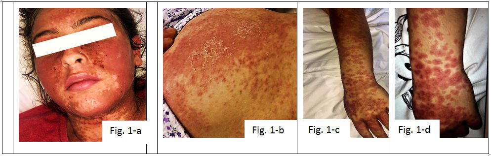 |
Figure 1: Clinical presentation of the first twin showing widespread body lesions. Some areas such as the face and the abdominal part (a and b) showed vesicle formation with skin sloughing |
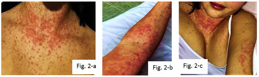 |
Figure 2: The second twin exhibited sparse macular lesions over the trunk area and arms. No mucosal involvement was noted |
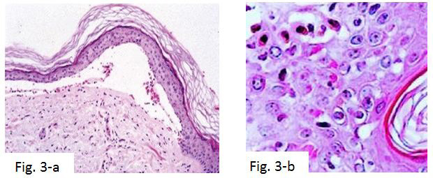 |
Figure 3: Light microscopy findings in the H&E-stained slides from a lesion of Stevens-Johnson syndrome showing single-cell necrosis extending to partial epidermal necrosis, extensive subepidermal blistering and sparse perivascular infiltration of lymphocytes and histiocytes |


